
Introduction
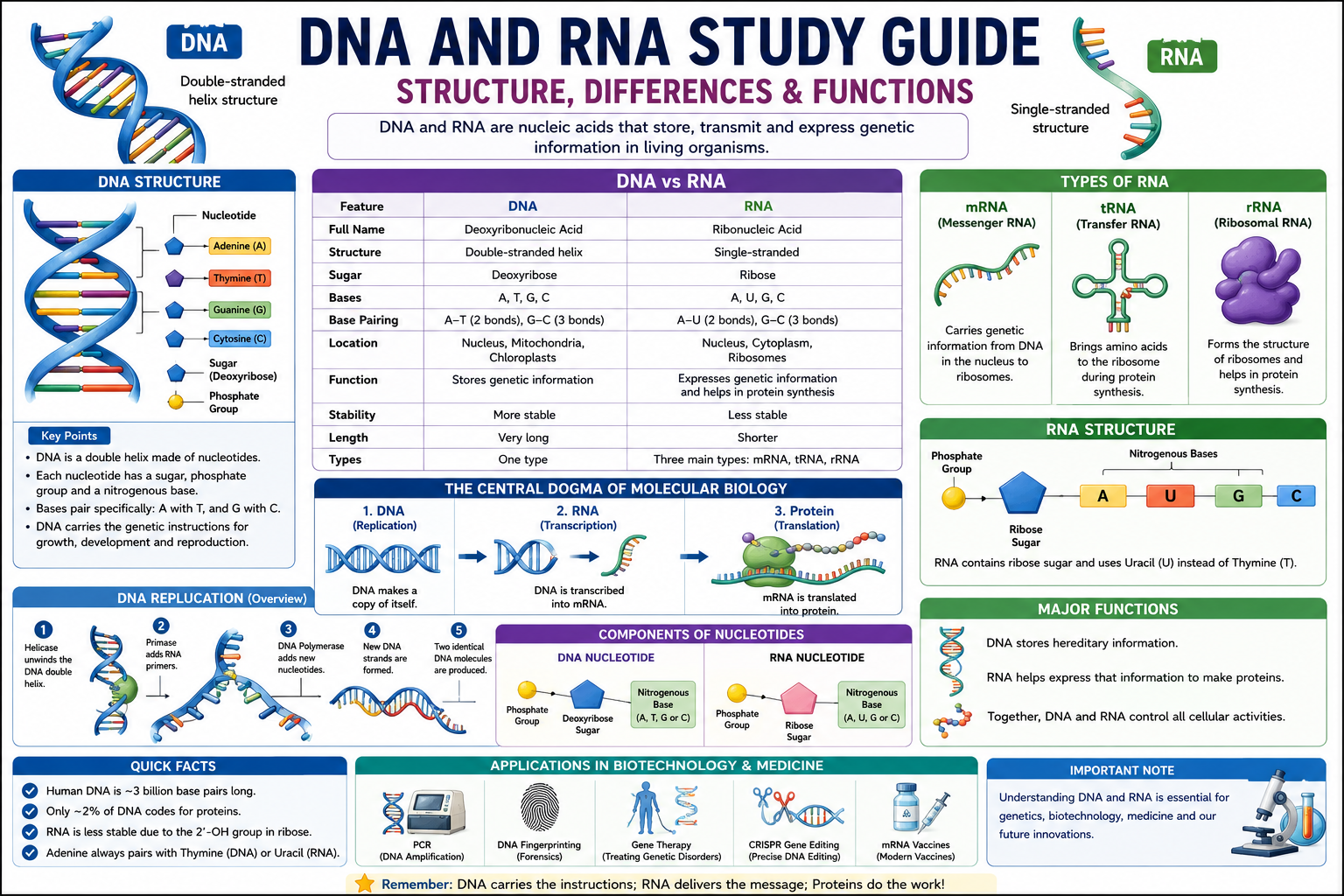
Inside almost every cell of your body, there’s a molecule so long that if you stretched it out fully, it would reach from here to the sun and back—several times over. That molecule is DNA, and it contains the complete instructions for building and running every living thing on Earth. Alongside it works RNA, a versatile molecule that reads those instructions, carries them to the protein-building machinery, and helps execute them with remarkable precision.
This DNA and RNA study guide is built to give you a thorough, clear, and genuinely useful understanding of both molecules—their structures, their functions, how they work together, and why understanding them is one of the most important things you can do in molecular biology. Whether you’re preparing for AP Biology, tackling a genetics unit in college, studying for medical entrance exams, or simply trying to understand what CRISPR actually does and why the world is excited about it, this guide has you covered.
Here’s the approach we’ll take: before memorizing any details, understand the logic. DNA stores information. RNA carries and uses that information. Proteins do the work. That central dogma of molecular biology—DNA → RNA → Protein—is the thread that runs through everything in this guide. Once you understand why each molecule exists and what job it does, the structural details become much easier to remember.
Over the next several thousand words, we’ll cover DNA and RNA structure in detail, walk through replication, transcription, and translation step by step, explore mutations and their consequences, examine how DNA technology is transforming medicine, and give you practice questions, a revision checklist, and everything else you need to walk into any exam feeling genuinely prepared.
Let’s start with the molecule that started everything.
Key Takeaways
By the end of this guide, you’ll be able to:
- Describe the structure of DNA including the double helix, nucleotides, base pairing, and the sugar-phosphate backbone
- Explain the structure and three major types of RNA (mRNA, tRNA, rRNA)
- Compare DNA and RNA across seven major features
- Describe DNA replication including the key enzymes and the semiconservative mechanism
- Explain transcription (DNA → mRNA) and translation (mRNA → protein) in sequential detail
- Define the genetic code and explain how codons specify amino acids
- Classify and explain the effects of point mutations, insertions, deletions, and chromosomal mutations
- Describe five major DNA technologies: PCR, DNA fingerprinting, genetic engineering, gene therapy, and CRISPR
- Avoid the most common exam mistakes in DNA and RNA questions
What Are DNA and RNA?
DNA (deoxyribonucleic acid) and RNA (ribonucleic acid) are nucleic acids—the category of biological macromolecules responsible for storing, transmitting, and using genetic information.
DNA is the master blueprint. It stores the hereditary information that determines the characteristics of every living organism—from the color of your eyes to the enzymes in your liver cells to the precise folding of proteins your immune system uses to fight disease. DNA is stable, double-stranded, and designed for long-term information storage. It stays safely in the nucleus (or nucleoid in prokaryotes) and rarely leaves.
RNA is the working copy. Because DNA is too precious to risk sending out into the bustling, enzyme-filled cytoplasm, the cell makes temporary RNA copies of specific genes when those genes need to be expressed. RNA carries the information from DNA to the ribosomes, where proteins are built.
Here’s an analogy that works well: DNA is the original architectural blueprint, locked safely in the architect’s office. RNA is the working copy taken to the construction site—disposable, temporary, but functionally precise.
Both DNA and RNA are polymers—long molecules made by linking smaller subunits called nucleotides. Despite their shared basic structure, they differ in important ways: in their sugar component, in one of their nitrogenous bases, in whether they’re single or double-stranded, and in their location and function in the cell.
Why DNA and RNA Are Important
Understanding DNA and RNA is not just an academic exercise. These molecules are at the center of medicine, agriculture, forensics, evolution, and virtually every area of modern biology.
Heredity and Inheritance
DNA carries the genetic information passed from parents to offspring. Every trait you inherited—your height, blood type, susceptibility to certain diseases, even some aspects of personality—traces back to DNA sequences passed down through generations.
Disease and Medicine
Many diseases—including cancer, genetic disorders, and viral infections—involve disruptions to DNA or RNA. Cancer results from mutations in DNA that disrupt normal cell cycle regulation. HIV is an RNA virus that integrates its genetic material into human DNA. Understanding these mechanisms is foundational to developing treatments.
Biotechnology
Every major advance in modern biotechnology involves manipulating DNA or RNA: PCR (used in COVID testing), DNA fingerprinting (forensic identification), CRISPR gene editing (potential cures for genetic diseases), mRNA vaccines (COVID-19 immunization), and recombinant DNA technology (insulin production).
Evolution
Mutations in DNA are the source of heritable variation that natural selection acts upon. Understanding DNA is understanding the molecular basis of evolution.
Forensics and Legal Justice
DNA profiling has transformed forensic science—exonerating the wrongly convicted and providing definitive evidence in criminal investigations.
History of DNA and RNA Discovery
The story of how we came to understand DNA and RNA is one of the most dramatic in the history of science—full of competition, overlooked contributions, and eureka moments.
Friedrich Miescher (1869)
Swiss physician Friedrich Miescher was the first to isolate nucleic acids—he called them “nuclein”—from white blood cells in pus from surgical bandages. He recognized these molecules were distinct from proteins, but their function remained mysterious for decades.
Phoebus Levene (early 1900s)
Levene identified the basic components of nucleotides—a phosphate group, a sugar, and a nitrogenous base—and distinguished DNA (containing deoxyribose) from RNA (containing ribose). He incorrectly proposed that DNA had a simple repeating pattern, which led many scientists to dismiss it as too simple to carry genetic information.
Frederick Griffith (1928)
Griffith’s famous transformation experiment with Streptococcus pneumoniae bacteria showed that something from heat-killed virulent bacteria could “transform” non-virulent bacteria into virulent ones. He didn’t know what the transforming substance was.
Oswald Avery, Colin MacLeod, and Maclyn McCarty (1944)
Building on Griffith’s work, this team demonstrated that the transforming substance was DNA—not protein, as many scientists expected. This was the first strong evidence that DNA carries genetic information, though it was met with significant skepticism.
Alfred Hershey and Martha Chase (1952)
Using bacteriophages (viruses that infect bacteria) and radioactive labels, Hershey and Chase conclusively demonstrated that it is DNA—not protein—that is injected into bacteria during infection, confirming DNA as the genetic material.
Erwin Chargaff (1950–1951)
Chargaff made a crucial observation: in any DNA sample, the amount of adenine always equals the amount of thymine, and the amount of guanine always equals the amount of cytosine (Chargaff’s rules). This suggested specific base pairing—a critical clue for the double helix structure.
Rosalind Franklin and Raymond Gosling (1952)
Franklin’s X-ray crystallography work produced “Photo 51″—a crucial X-ray diffraction image of DNA that revealed its helical structure and key dimensions. This data was shared with Watson and Crick without Franklin’s knowledge, a deeply controversial aspect of the story.
James Watson and Francis Crick (1953)
Using Franklin’s data along with other evidence, Watson and Crick built their famous molecular model and published the double helix structure of DNA in Nature in April 1953. Watson, Crick, and Maurice Wilkins received the Nobel Prize in Physiology or Medicine in 1962. Franklin, who died in 1958, was not eligible.
DNA Structure Explained
Double Helix Structure
DNA exists as a double helix—two complementary strands of nucleotides wound around each other in a spiral, like a twisted ladder. Watson and Crick’s model elegantly explained both how DNA stores information (in the sequence of bases) and how it can be copied (each strand serves as a template for the other).
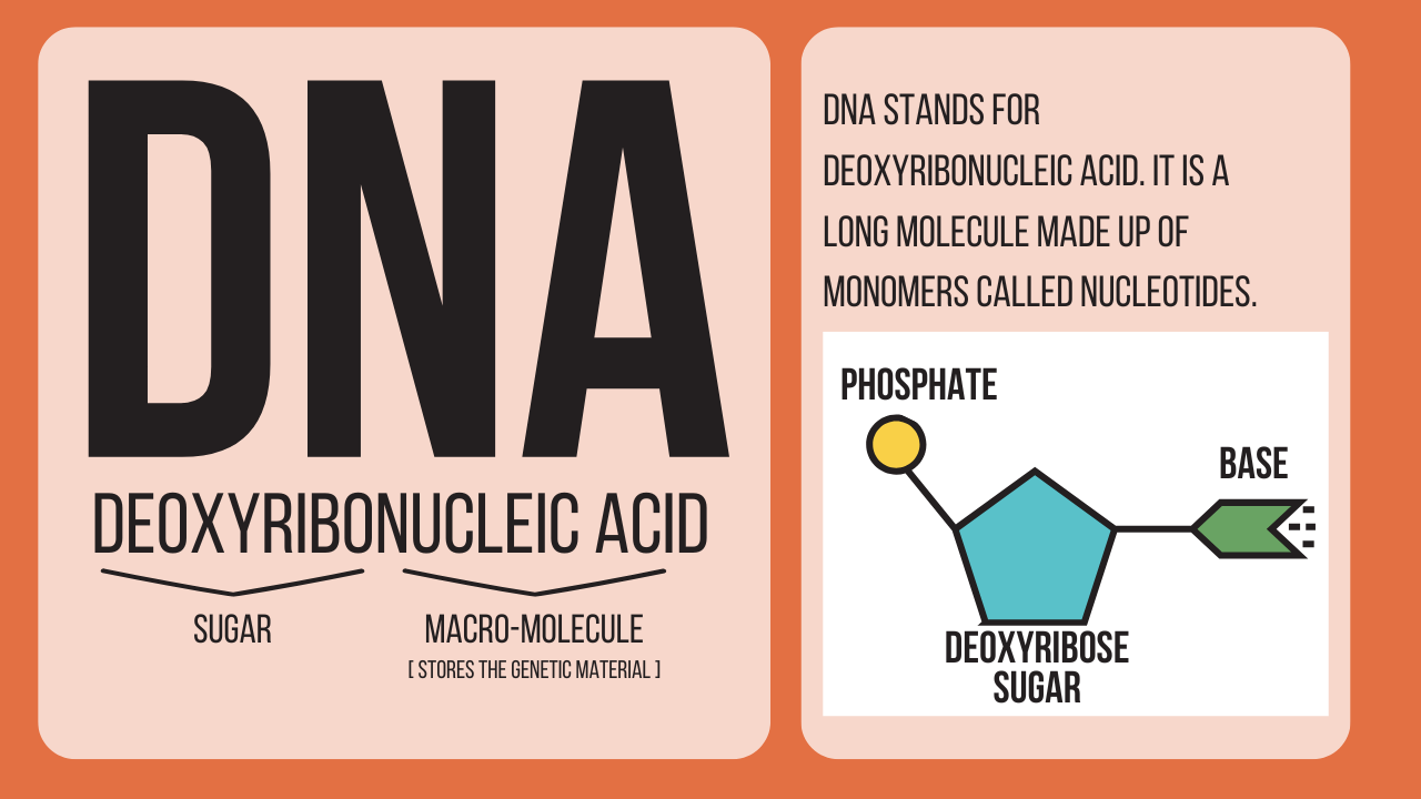
The double helix has several key physical features:
- Diameter: Approximately 2 nanometers
- Pitch: One complete turn every 10 base pairs (~3.4 nm)
- Antiparallel orientation: The two strands run in opposite directions—one 5’→3′, the other 3’→5′. This antiparallel arrangement is essential for DNA replication and transcription.
Nucleotides
Each strand of DNA is a polymer of nucleotides—the monomers of nucleic acids. Each nucleotide consists of three components:
- A pentose (5-carbon) sugar – In DNA, this is deoxyribose (lacks an oxygen at the 2′ carbon position)
- A phosphate group – Negatively charged; links nucleotides together in the backbone
- A nitrogenous base – The information-carrying component; one of four types
Nucleotides are linked together by phosphodiester bonds between the 3′ carbon of one sugar and the 5′ carbon of the next, forming the continuous sugar-phosphate backbone. The bases project inward, like the rungs of a ladder.
Nitrogenous Bases
DNA contains four nitrogenous bases, divided into two categories:
Purines (double-ring structures):
- Adenine (A)
- Guanine (G)
Pyrimidines (single-ring structures):
- Cytosine (C)
- Thymine (T)
Base pairing rules (Chargaff’s rules):
- Adenine (A) pairs with Thymine (T) — joined by 2 hydrogen bonds
- Guanine (G) pairs with Cytosine (C) — joined by 3 hydrogen bonds
The G-C pair is stronger (3 hydrogen bonds vs 2) which is why DNA regions with high G-C content are more thermally stable and harder to separate. This has practical implications in PCR and molecular biology techniques.
Memory aid: “Pure As Gold” — Purines are Adenine and Guanine. “CUT the PyrimidiNE” — Pyrimidines are Cytosine, Uracil (in RNA), and Thymine.
Sugar-Phosphate Backbone
The sugar-phosphate backbone forms the structural framework of each DNA strand. The alternating deoxyribose sugars and phosphate groups are linked by covalent phosphodiester bonds—creating a strong, stable backbone. The negatively charged phosphate groups make DNA an acidic molecule (hence the “acid” in nucleic acid) and are responsible for its negative charge in gel electrophoresis.
The backbone runs in a specific direction. The 5′ end has a free phosphate group; the 3′ end has a free hydroxyl (−OH) group. New nucleotides are always added to the 3′ end during replication and transcription—the polymerases work 5′ to 3′.
Hydrogen Bonds
The two strands of the double helix are held together by hydrogen bonds between complementary base pairs. While individual hydrogen bonds are relatively weak, the cumulative effect of thousands of hydrogen bonds along a DNA molecule provides substantial stability. Crucially, hydrogen bonds can also be broken—temporarily, during replication and transcription—which is essential for accessing the information encoded in the base sequence.
The specific base pairing enforced by hydrogen bonds means that once you know the sequence of one strand, you automatically know the sequence of the other—complementarity. This is what makes accurate DNA replication possible.
RNA Structure Explained
Single-Stranded Structure
Unlike DNA, RNA is typically single-stranded. This single-stranded nature is not a limitation—it’s a feature that allows RNA molecules to fold back on themselves, forming complex three-dimensional shapes through intramolecular base pairing. These shapes are critical for tRNA and rRNA function.
Ribose Sugar
The sugar in RNA is ribose—it has a hydroxyl group (−OH) at the 2′ carbon position, unlike deoxyribose which has just a hydrogen (−H) there. This seemingly small difference has significant consequences:
- RNA is less chemically stable than DNA (the 2’−OH group makes it more susceptible to hydrolysis)
- RNA’s instability is actually advantageous—it means RNA messages can be degraded after use, giving the cell fine control over gene expression
- DNA’s greater stability makes it ideal for long-term information storage
Nitrogenous Bases
RNA contains the same bases as DNA except that thymine (T) is replaced by uracil (U). Like thymine, uracil pairs with adenine—but uracil lacks the methyl group on thymine and is cheaper for the cell to synthesize.
RNA bases:
- Adenine (A)
- Guanine (G)
- Cytosine (C)
- Uracil (U) — replaces Thymine
Messenger RNA (mRNA)
mRNA is the information-carrying form of RNA—the “message” that carries genetic information from DNA in the nucleus to ribosomes in the cytoplasm.
Key features:
- Linear, single-stranded
- Carries the genetic code in sequences of three-nucleotide codons
- Produced during transcription from a DNA template
- Relatively short-lived—degraded after translation (half-life of minutes to hours in prokaryotes; hours to days in eukaryotes)
- In eukaryotes, pre-mRNA undergoes processing: 5′ cap addition, poly-A tail addition, and splicing out of introns before leaving the nucleus
Each codon in the mRNA sequence specifies a particular amino acid (or a start/stop signal) during translation. The sequence of codons in mRNA thus determines the sequence of amino acids in the resulting protein.
Transfer RNA (tRNA)
tRNA is the adaptor molecule that decodes the mRNA message—bridging the genetic code and protein synthesis by matching codons to amino acids.
Key features:
- ~75–95 nucleotides long
- Folds into a characteristic cloverleaf secondary structure (due to intramolecular base pairing) and an L-shaped tertiary structure
- Each tRNA has two critical regions:
- Anticodon loop – Contains the three-nucleotide anticodon that base-pairs with the complementary mRNA codon
- 3′ acceptor stem – The site where a specific amino acid is attached (by an enzyme called aminoacyl-tRNA synthetase)
- There is at least one tRNA for each amino acid; the cell contains over 40 different tRNA species
Ribosomal RNA (rRNA)
rRNA is a structural and catalytic component of ribosomes—the molecular machines where protein synthesis occurs.
Key features:
- Most abundant RNA in the cell (~80% of total cellular RNA)
- Combines with ribosomal proteins to form the large and small subunits of ribosomes
- The large subunit’s rRNA has catalytic activity—it’s the component that actually forms peptide bonds between amino acids (making the ribosome a ribozyme—an RNA molecule with enzymatic activity)
- In prokaryotes: 23S, 16S, and 5S rRNAs; in eukaryotes: 28S, 18S, 5.8S, and 5S rRNAs
The 16S rRNA in prokaryotes is used in phylogenetic studies to classify bacteria—its sequence is conserved enough to be compared across species but variable enough to reveal evolutionary relationships.
DNA vs RNA Comparison Table
| Feature | DNA | RNA |
|---|---|---|
| Full name | Deoxyribonucleic acid | Ribonucleic acid |
| Sugar | Deoxyribose (no 2′-OH) | Ribose (has 2′-OH) |
| Strands | Double-stranded | Single-stranded (usually) |
| Bases | A, T, G, C | A, U, G, C |
| Base replacing T/U | Thymine (T) | Uracil (U) |
| Location | Nucleus, mitochondria, chloroplasts | Nucleus, cytoplasm, ribosomes |
| Stability | Very stable (long-term storage) | Less stable (temporary) |
| Function | Stores genetic information | Carries, interprets, uses genetic info |
| Types | One primary type | mRNA, tRNA, rRNA (+ others) |
| Length | Very long (millions of base pairs) | Shorter (hundreds to thousands of bases) |
| Role in evolution | Source of heritable mutations | Can be genetic material in RNA viruses |
Components of DNA and RNA
Both DNA and RNA are built from nucleotides, but their components differ in key ways:
| Component | DNA Nucleotide | RNA Nucleotide |
|---|---|---|
| Sugar | Deoxyribose | Ribose |
| Phosphate | Yes (1–3 phosphates) | Yes (1–3 phosphates) |
| Purine bases | Adenine, Guanine | Adenine, Guanine |
| Pyrimidine bases | Cytosine, Thymine | Cytosine, Uracil |
| Bonds between nucleotides | Phosphodiester bonds | Phosphodiester bonds |
| Base pairing | A-T (2 H bonds), G-C (3 H bonds) | A-U (2 H bonds), G-C (3 H bonds) |
DNA Replication Explained
DNA replication is the process by which a cell copies its entire genome before cell division, ensuring each daughter cell receives a complete, accurate copy of the genetic information.
The mechanism is semiconservative: each new double helix consists of one original (parental) strand and one newly synthesized strand. This was confirmed by the elegant Meselson-Stahl experiment (1958) using nitrogen isotope labeling.
Key enzymes and proteins in DNA replication:
| Protein/Enzyme | Function |
|---|---|
| Helicase | Unwinds and separates the double helix by breaking hydrogen bonds |
| Primase | Synthesizes short RNA primers to start replication |
| DNA Polymerase III (prokaryotes) | Adds new nucleotides 5’→3′; proofreads |
| DNA Polymerase I | Removes RNA primers; fills gaps with DNA |
| DNA Ligase | Joins Okazaki fragments; seals nicks in DNA |
| Single-strand binding proteins (SSBPs) | Stabilize unwound single strands |
| Topoisomerase | Relieves tension ahead of the replication fork |
Replication process step by step:
- Initiation – Replication begins at specific sequences called origins of replication (prokaryotes have one; eukaryotes have thousands, allowing faster replication of large genomes). Helicase binds and unwinds the double helix, creating a replication fork.
- Priming – Primase synthesizes short RNA primers (8–12 nucleotides) complementary to the template strand. DNA polymerase cannot start a new strand from scratch—it can only extend an existing one, so primers are essential.
- Elongation – DNA polymerase adds nucleotides 5’→3′ to the growing strand, reading the template 3’→5′. Because of antiparallel orientation:
- The leading strand is synthesized continuously toward the replication fork
- The lagging strand is synthesized discontinuously, away from the fork, in short fragments called Okazaki fragments
- Primer removal and gap filling – RNA primers are removed by DNA Polymerase I, which fills the gaps with DNA nucleotides.
- Ligation – DNA Ligase seals the nicks between adjacent DNA fragments, creating a continuous strand.
- Termination – Replication ends when the forks meet (in circular bacterial chromosomes) or when the ends of chromosomes are reached (in linear eukaryotic chromosomes—where telomeres protect chromosome ends from shortening).
Replication fidelity: DNA Polymerase proofreads as it goes, detecting and correcting mismatched bases. The error rate is approximately 1 in 10 billion nucleotides after proofreading—an extraordinary level of accuracy.
Transcription (DNA to RNA)
Transcription is the process by which the information in a DNA sequence is copied into a complementary RNA sequence. It’s the first step in gene expression—the process of converting genetic information into functional proteins.
Key participants:
- RNA polymerase – The enzyme that synthesizes RNA (unlike DNA polymerase, RNA polymerase doesn’t need a primer)
- Template strand – The strand of DNA read by RNA polymerase (3’→5′)
- Coding strand (non-template strand) – Has the same sequence as the resulting mRNA (except T is replaced by U)
- Promoter – DNA sequence where RNA polymerase binds to initiate transcription; in eukaryotes, often includes the TATA box
- Transcription factors – Proteins that help RNA polymerase bind to promoters
Transcription steps:
1. Initiation
RNA polymerase (with associated transcription factors in eukaryotes) binds to the promoter sequence upstream of the gene. The double helix is locally unwound, exposing the template strand.
2. Elongation
RNA polymerase moves along the template strand 3’→5′, synthesizing RNA 5’→3′ by adding ribonucleotides complementary to the template. The RNA strand grows as the polymerase moves through the gene.
3. Termination
Transcription ends when RNA polymerase reaches a terminator sequence. In prokaryotes, this can cause the RNA to fold into a hairpin structure that stalls polymerase. In eukaryotes, termination is more complex and involves cleavage of the RNA molecule.
Eukaryotic pre-mRNA processing:
Before the primary transcript (pre-mRNA) can leave the nucleus, it undergoes extensive processing:
- 5′ capping – A modified guanosine cap is added to the 5′ end; protects mRNA from degradation and assists ribosome binding
- Poly-A tail – ~150–250 adenine nucleotides are added to the 3′ end; protects from degradation and assists nuclear export
- Splicing – Introns (non-coding sequences) are removed by the spliceosome (a complex of snRNAs and proteins); exons (coding sequences) are joined together
The mature mRNA is then exported through nuclear pores to the cytoplasm for translation.
Important Fact: The discovery of introns and splicing (by Roberts and Sharp in 1977, for which they received the Nobel Prize) was revolutionary—it revealed that most eukaryotic genes are not continuous coding sequences. A single pre-mRNA can be alternatively spliced to produce different proteins from the same gene—this is called alternative splicing and is one reason humans have far more protein diversity than our gene count would suggest.
Translation (RNA to Protein)
Translation is the process by which the mRNA sequence is decoded to synthesize a specific polypeptide chain (protein). It occurs at ribosomes—molecular machines consisting of rRNA and protein.
Ribosomes:
- Consist of a small subunit (reads mRNA) and a large subunit (catalyzes peptide bond formation)
- Have three tRNA-binding sites:
- A site (aminoacyl site) – Where incoming aminoacyl-tRNA binds
- P site (peptidyl site) – Where the growing polypeptide chain is held
- E site (exit site) – Where the empty tRNA exits
Translation steps:
1. Initiation
- The small ribosomal subunit binds to the mRNA at the 5′ cap (eukaryotes) or Shine-Dalgarno sequence (prokaryotes)
- It scans for the start codon: AUG (codes for methionine)
- The initiator tRNA (carrying methionine) binds the start codon at the P site
- The large subunit joins to complete the ribosome
2. Elongation
This is where the actual protein chain grows, one amino acid at a time:
| Step | What Happens |
|---|---|
| Codon recognition | An aminoacyl-tRNA with the correct anticodon enters the A site |
| Peptide bond formation | The ribosome (specifically the peptidyl transferase of rRNA) catalyzes formation of a peptide bond between the amino acids |
| Translocation | The ribosome moves one codon (3 nucleotides) along the mRNA in the 5’→3′ direction; the tRNA in P site moves to E site and exits; A site tRNA moves to P site; A site becomes empty for next aminoacyl-tRNA |
3. Termination
Elongation continues until a stop codon (UAA, UAG, or UGA) enters the A site. Stop codons are not recognized by any tRNA—instead, release factors (proteins) bind the stop codon, triggering release of the completed polypeptide chain. The ribosome then dissociates from the mRNA.
Post-translational modification:
The newly synthesized polypeptide is not yet a functional protein. It typically undergoes:
- Folding (often assisted by chaperone proteins)
- Cleavage of the initiator methionine
- Chemical modifications (phosphorylation, glycosylation, acetylation)
- Transport to the correct cellular location
Gene Expression Explained
Gene expression is the full process by which the information encoded in a gene is used to produce a functional product—most commonly a protein. It encompasses transcription, RNA processing, translation, and post-translational modification.
The Central Dogma of Molecular Biology (Francis Crick, 1958):
DNA → RNA → ProteinThis directional flow is the foundational principle of molecular biology. Information flows from DNA to RNA to protein—not in reverse (with some exceptions: retroviruses like HIV use reverse transcriptase to convert RNA back to DNA).
Gene regulation is the control of when, where, and how much of a gene product is made. Cells in your body all contain the same DNA, but different cells express different genes—which is why a liver cell looks and functions differently from a neuron, despite having identical DNA.
Gene expression is regulated at multiple levels:
- Transcriptional regulation – Transcription factors, enhancers, silencers, promoter accessibility
- Post-transcriptional regulation – mRNA stability, alternative splicing, RNA interference (miRNA, siRNA)
- Translational regulation – Ribosome binding, initiation factors
- Post-translational regulation – Protein modification, degradation, localization
Genetic Code and Codons
The genetic code is the set of rules by which nucleotide sequences in mRNA are translated into amino acid sequences in proteins. It’s essentially the molecular “dictionary” of life.
Key features of the genetic code:
- Triplet code – Three consecutive nucleotides (a codon) specify one amino acid. With 4 possible nucleotides at each of 3 positions, there are 4³ = 64 possible codons.
- 64 codons; 20 amino acids – Most amino acids are specified by more than one codon (degeneracy/redundancy)
- Start codon: AUG – Codes for methionine; signals the start of translation
- Stop codons: UAA, UAG, UGA – Do not code for any amino acid; signal termination of translation
- Universal – The same genetic code is used by virtually all organisms on Earth (with minor exceptions), which is compelling evidence for the common ancestry of all life
- Non-overlapping – Each nucleotide is part of only one codon
- Unambiguous – Each codon codes for only one amino acid (though one amino acid can be coded by multiple codons)
Examples of codons:
| Codon | Amino Acid |
|---|---|
| AUG | Methionine (Start) |
| UUU / UUC | Phenylalanine |
| GGG / GGC / GGA / GGU | Glycine |
| UAA / UAG / UGA | Stop |
| GAA / GAG | Glutamic acid |
Mutations and Their Effects
A mutation is any permanent change in the DNA sequence of an organism. Mutations are the source of genetic variation—they can be neutral, beneficial, or harmful depending on how they affect protein structure and function.
Point Mutations
Point mutations involve a change in a single nucleotide base pair.
Types of point mutations:
- Silent (synonymous) mutation – The base change results in a different codon that still codes for the same amino acid (due to degeneracy of the genetic code). No change in protein; often harmless. Example: GGA → GGU, both code for glycine.
- Missense mutation – The base change results in a codon that codes for a different amino acid. The protein may be functional, partially functional, or non-functional depending on the amino acid’s importance. Example: Sickle cell anemia—a single A→T change converts GAG (glutamic acid) to GUG (valine) in the hemoglobin beta chain, causing the protein to misfold.
- Nonsense mutation – The base change creates a premature stop codon, truncating the protein. Usually results in a non-functional protein. Example: Cystic fibrosis can be caused by nonsense mutations in the CFTR gene.
Insertions
An insertion adds one or more nucleotides to the DNA sequence. Unless the number of nucleotides inserted is a multiple of 3, insertions cause a frameshift mutation—shifting the reading frame so that all downstream codons are read differently. Frameshifts usually produce a severely altered, non-functional protein.
Deletions
A deletion removes one or more nucleotides. Like insertions, deletions that are not multiples of 3 cause frameshift mutations with cascading effects on all downstream codons. Example: Duchenne muscular dystrophy is often caused by frameshift deletions in the dystrophin gene.
Chromosomal Mutations
Chromosomal mutations involve large-scale changes in chromosome structure or number:
- Deletion – A chromosomal segment is lost
- Duplication – A segment is duplicated
- Inversion – A segment is reversed
- Translocation – A segment moves to a different chromosome
- Aneuploidy – Incorrect chromosome number (e.g., trisomy 21/Down syndrome = extra chromosome 21)
- Polyploidy – Multiplication of entire chromosome sets (common in plants; rare in animals)
Causes of mutations:
- Spontaneous errors during DNA replication (despite proofreading)
- Mutagens: UV radiation (causes thymine dimers), X-rays, chemical mutagens (e.g., aflatoxin, nitrous acid), certain viruses
DNA Packaging into Chromosomes
The human genome contains approximately 3.2 billion base pairs of DNA. If fully extended, the DNA in a single human cell would be about 2 meters long. Yet it must fit into a nucleus approximately 6 micrometers in diameter. The solution is extraordinary hierarchical packaging.
Levels of DNA packaging:
- DNA + Histones → Nucleosomes
- DNA wraps approximately 1.7 times around an octamer of histone proteins to form a nucleosome (the fundamental unit of chromatin)
- Nucleosomes are connected by linker DNA, giving the appearance of “beads on a string”
- Nucleosome arrays → 30 nm fiber
- Nucleosome strings fold into a more compact 30 nm chromatin fiber (exact structure debated)
- 30 nm fiber → Loops
- The fiber forms looped domains attached to a protein scaffold
- Loops → Coiled coils → Condensed chromosome
- During cell division, chromatin is maximally compacted into the visible chromosome structure
Euchromatin vs Heterochromatin:
- Euchromatin – Loosely packed; transcriptionally active; genes can be expressed
- Heterochromatin – Tightly packed; transcriptionally silent; includes centromeres and telomeres
The packaging state of DNA profoundly affects gene expression—genes in tightly packaged regions are inaccessible to transcription machinery and therefore silenced. This is the basis of epigenetic regulation.
Role of DNA and RNA in Protein Synthesis
Protein synthesis is the central function that DNA and RNA cooperate to accomplish. Here’s how all three nucleic acid types work together:
The complete flow:
DNA (nucleus)
    ↓ Transcription (RNA polymerase)
pre-mRNA (nucleus)
    ↓ Processing (capping, poly-A tail, splicing)
mature mRNA (nucleus → cytoplasm)
    ↓ Translation (ribosome + tRNA)
Polypeptide chain
    ↓ Post-translational modification
Functional protein
Each molecule’s role:
| Molecule | Role in Protein Synthesis |
|---|---|
| DNA | Provides the template for transcription; contains the gene sequence |
| mRNA | Carries the genetic message from nucleus to ribosome; contains codons |
| tRNA | Brings specific amino acids to the ribosome; matches codons via anticodon |
| rRNA | Structural and catalytic component of ribosome; catalyzes peptide bond formation |
| RNA polymerase | Synthesizes mRNA during transcription |
| Ribosome | Platform where translation occurs; reads mRNA and builds polypeptide |
DNA Technology and Biotechnology
Understanding DNA and RNA has enabled a technological revolution that is transforming medicine, agriculture, forensics, and basic research.
PCR (Polymerase Chain Reaction)
PCR is a technique for amplifying specific DNA sequences—making millions of copies from a tiny amount of starting material. Invented by Kary Mullis in 1983 (Nobel Prize 1993), PCR has become one of the most widely used tools in molecular biology.
How PCR works:
- Denaturation (~95°C) – DNA strands are separated by heat
- Annealing (~50–65°C) – Specific primers bind to their complementary sequences flanking the target region
- Extension (~72°C) – Taq polymerase (a heat-stable DNA polymerase from Thermus aquaticus) extends the primers, copying the target sequence
Each cycle doubles the amount of target DNA. After 30 cycles: over one billion copies from a single DNA molecule.
Applications: COVID-19 diagnostic testing, forensic DNA analysis, paternity testing, detecting genetic mutations, cloning genes, ancient DNA analysis.
DNA Fingerprinting
DNA fingerprinting (DNA profiling) uses specific variable regions of the genome—particularly short tandem repeats (STRs)—to create a unique genetic profile for an individual. The probability of two unrelated individuals having identical STR profiles at multiple loci is vanishingly small.
Applications: Forensic identification (crime scenes, missing persons), paternity and relationship testing, identifying victims of disasters, wildlife conservation genetics.
Genetic Engineering
Genetic engineering involves deliberately modifying an organism’s genome by inserting, deleting, or altering genes. The basic toolkit includes:
- Restriction endonucleases – Bacterial enzymes that cut DNA at specific sequences (“molecular scissors”)
- Recombinant DNA technology – Combining DNA from different sources, typically by inserting a gene of interest into a plasmid vector
- Expression systems – Using bacteria, yeast, or mammalian cells to produce proteins from inserted genes
Applications: Producing human insulin in bacteria, developing herbicide-resistant crops (Bt corn, Roundup Ready soybeans), producing vaccines, manufacturing industrial enzymes.
Gene Therapy
Gene therapy involves introducing genetic material into a patient’s cells to treat or prevent disease. It holds particular promise for genetic disorders caused by single-gene mutations.
Approaches:
- Somatic gene therapy – Modifying non-reproductive cells; changes affect only the patient, not their offspring
- Germline gene therapy – Modifying embryonic cells; changes would be inherited by future generations (highly controversial and banned in most countries)
Delivery methods:
- Viral vectors – Modified viruses (adenoviruses, AAVs, lentiviruses) that can deliver DNA into cells
- Lipid nanoparticles – Used to deliver mRNA (as in mRNA vaccines)
- Direct injection of DNA or RNA
Approved therapies: Luxturna (for a form of inherited blindness), Zolgensma (for spinal muscular atrophy—the most expensive drug ever developed), and many others in clinical trials.
CRISPR Gene Editing
CRISPR-Cas9 is the most significant advance in genetic engineering since recombinant DNA technology. Derived from a bacterial immune system, CRISPR (Clustered Regularly Interspaced Short Palindromic Repeats) allows scientists to cut DNA at any specific location in the genome with unprecedented precision.
How CRISPR works:
- A guide RNA (gRNA) is designed to match the target DNA sequence
- The gRNA directs Cas9 (a nuclease enzyme) to the target location
- Cas9 cuts both strands of DNA at the target
- The cell’s own repair mechanisms fix the break:
- Non-homologous end joining (NHEJ) – Error-prone repair; often creates small insertions or deletions that knock out the gene
- Homology-directed repair (HDR) – If a repair template is provided, the desired sequence can be precisely inserted
Applications: Potential cures for sickle cell disease and beta-thalassemia (clinical trials showing remarkable success), developing disease-resistant crops, research tools for understanding gene function, potential HIV treatment.
Important Fact: In 2020, Jennifer Doudna and Emmanuelle Charpentier received the Nobel Prize in Chemistry for developing CRISPR-Cas9 gene editing—the first Nobel awarded for a technology with such immediate and direct medical applications.
DNA and RNA in Human Health
Genetic Disorders
Many human diseases arise from DNA mutations:
- Sickle cell anemia – Missense mutation in hemoglobin beta gene
- Cystic fibrosis – Most commonly a 3-nucleotide deletion (ΔF508) in the CFTR gene
- Huntington’s disease – Expansion of a trinucleotide repeat (CAG) in the huntingtin gene
- Fragile X syndrome – Expansion of CGG repeats in the FMR1 gene
- Down syndrome – Trisomy 21 (chromosomal mutation)
Cancer
Cancer is fundamentally a disease of gene expression gone wrong. Mutations in:
- Proto-oncogenes → Oncogenes (accelerate cell division)
- Tumor suppressor genes (e.g., p53, BRCA1) → Loss of function (remove brakes on cell division)
- DNA repair genes → Accumulation of additional mutations
Viral Diseases
Many viruses use RNA as their genetic material:
- HIV – RNA retrovirus; uses reverse transcriptase to convert RNA genome into DNA
- SARS-CoV-2 – RNA virus; its RNA genome is directly translated upon cell entry
- Influenza – RNA virus with a segmented genome; the segmentation allows genetic reassortment, contributing to new strains
mRNA Vaccines
The COVID-19 pandemic brought mRNA technology to global attention. mRNA vaccines introduce mRNA encoding a viral protein (like the spike protein of SARS-CoV-2) into cells. Ribosomes translate the mRNA into the viral protein, which stimulates an immune response. The mRNA is degraded after serving its purpose—it never enters the nucleus and cannot affect DNA.
Common DNA and RNA Terms Every Student Should Know
| Term | Definition |
|---|---|
| Nucleotide | Monomer of nucleic acids; consists of a phosphate, sugar, and nitrogenous base |
| Double helix | The coiled, ladder-like structure of DNA |
| Base pair | Complementary bases joined by hydrogen bonds (A-T, G-C in DNA; A-U, G-C in RNA) |
| Antiparallel | The two strands of DNA running in opposite directions (5’→3′ and 3’→5′) |
| Semiconservative replication | Each new DNA double helix retains one original strand |
| Transcription | Synthesis of RNA from a DNA template |
| Translation | Synthesis of protein from an mRNA template |
| Codon | Three-nucleotide sequence in mRNA coding for an amino acid |
| Anticodon | Three-nucleotide sequence in tRNA complementary to a codon |
| Promoter | DNA sequence where RNA polymerase binds to initiate transcription |
| Intron | Non-coding sequence in eukaryotic pre-mRNA; removed during splicing |
| Exon | Coding sequence in eukaryotic pre-mRNA; retained in mature mRNA |
| Frameshift | Mutation caused by insertion or deletion of non-multiple-of-3 nucleotides |
| Gene | A sequence of DNA that encodes a functional product (usually a protein) |
| Genome | The complete DNA sequence of an organism |
| Chromatin | Complex of DNA and proteins (histones) in the eukaryotic nucleus |
| Nucleosome | DNA wrapped around histone octamer; fundamental unit of chromatin |
| Telomere | Repetitive DNA sequence at chromosome ends; protects against shortening |
| Central dogma | DNA → RNA → Protein; the directional flow of genetic information |
| Reverse transcriptase | Enzyme that synthesizes DNA from an RNA template (used by retroviruses) |
Common Mistakes Students Make
These are the errors that consistently appear in student responses on DNA and RNA topics.
1. Saying DNA contains uracil or RNA contains thymine
DNA contains thymine; RNA contains uracil. This seems basic but students frequently swap them under exam pressure. Memorize: RNA uses Uracil; DNA uses Thymine. The U/T distinction directly correlates with the ribose/deoxyribose sugar distinction.
2. Confusing the template strand with the coding strand
The template strand (also called the antisense or non-coding strand) is the one read by RNA polymerase—3’→5′. The coding strand (sense strand) has the same sequence as the mRNA (with T instead of U). Students often incorrectly state that mRNA is complementary to the coding strand—it’s complementary to the template strand but identical (in sequence) to the coding strand.
3. Confusing transcription and translation
Transcription = DNA → mRNA. Translation = mRNA → protein. These are different processes in different locations with different machinery. A reliable way to remember: Transcription produces transcript (RNA). Translation produces protein (because ribosomes translate the nucleotide language into the amino acid language).
4. Stating that replication is conservative (not semiconservative)
Replication is semiconservative: each new helix has one old strand and one new strand. Conservative replication (both old strands stay together, new helix is all new) was the alternative hypothesis before Meselson and Stahl’s experiment. If you write “conservative” on an exam, you’ll lose the mark.
5. Forgetting that the stop codons don’t code for any amino acid
UAA, UAG, and UGA are stop codons—they signal termination of translation. No tRNA has anticodons for these; release factors bind instead. Students sometimes write that stop codons “code for a stop amino acid,” which is incorrect.
6. Confusing introns and exons
Introns are removed (“introns go out”). Exons are expressed (“exons are expressed”). These simple memory cues—while imperfect biologically speaking—are reliable for exam purposes. Introns are non-coding; exons are coding sequences that appear in mature mRNA.
Best Tips to Study DNA and RNA
1. Learn the central dogma as your framework, then hang the details on it
Everything in DNA/RNA biology connects to DNA → RNA → Protein. Before memorizing enzymes, steps, and molecular details, make sure you understand the overall information flow. Every process you study either contributes to or regulates one step of this central dogma.
2. Draw, draw, draw
Sketch the double helix with labeled base pairs, hydrogen bonds, and sugar-phosphate backbone from memory. Draw the nucleosome. Draw the ribosome with A, P, and E sites. Draw the transcription bubble. Drawing forces active recall and reveals knowledge gaps faster than re-reading ever will.
3. Use base-pairing rules constantly
Practice writing complementary DNA and RNA sequences until it’s automatic. Given a DNA template: 3′-ATCGTA-5′, immediately know the mRNA: 5′-UAGCAU-3′. Then practice with tRNA anticodons. This is the kind of manipulation that appears in virtually every molecular biology exam.
4. Connect mutations to real diseases
Every major mutation type has a famous disease example: missense → sickle cell anemia; deletion frameshift → Duchenne muscular dystrophy; trinucleotide repeat expansion → Huntington’s disease; chromosomal trisomy → Down syndrome. Disease connections make abstract mutation types unforgettable.
5. Understand the logic of each enzyme’s role
Don’t just memorize that helicase unwinds DNA. Ask why the DNA needs to be unwound. Because polymerase needs single-stranded template to read. Ask why primase is needed. Because DNA polymerase can’t start from scratch. Understanding the why means you can reconstruct enzyme functions even if you blank on names during an exam.
6. Practice codon/anticodon problems
Work through several problems where you’re given a DNA sequence and asked to determine the mRNA sequence, the tRNA anticodon, and the amino acid sequence. These multi-step problems are common on exams and require fluency with all three molecular levels simultaneously.
DNA and RNA Practice Questions
20 Multiple Choice Questions with Answers
- Which sugar is found in DNA but NOT in RNA?
- A) Ribose
- B) Deoxyribose ✓
- C) Glucose
- D) Fructose
- Which nitrogenous base is found in RNA but NOT in DNA?
- A) Thymine
- B) Adenine
- C) Cytosine
- D) Uracil ✓
- Which base pairs with guanine in DNA?
- A) Adenine
- B) Thymine
- C) Cytosine ✓
- D) Uracil
- The semiconservative model of DNA replication means:
- A) Both strands are newly synthesized
- B) Each new double helix contains one original and one new strand ✓
- C) One new double helix is all new; one is all original
- D) Only half the DNA is replicated each cycle
- Which enzyme unwinds the DNA double helix during replication?
- A) DNA Polymerase
- B) Primase
- C) Ligase
- D) Helicase ✓
- During transcription, which molecule serves as the template?
- A) mRNA
- B) tRNA
- C) The template strand of DNA ✓
- D) The coding strand of DNA
- Which of the following is the start codon?
- A) UAA
- B) UGA
- C) AUG ✓
- D) UAG
- The anticodon is located on:
- A) mRNA
- B) rRNA
- C) tRNA ✓
- D) DNA
- Which type of mutation adds or removes nucleotides not in multiples of three?
- A) Silent mutation
- B) Missense mutation
- C) Frameshift mutation ✓
- D) Nonsense mutation
- Introns are:
- A) Coding sequences that appear in mature mRNA
- B) Non-coding sequences removed during splicing ✓
- C) Alternative start codons
- D) Regulatory sequences in the promoter
- Which type of RNA carries genetic information from the nucleus to ribosomes?
- A) tRNA
- B) mRNA ✓
- C) rRNA
- D) snRNA
- Which base pairing rule is correct for DNA?
- A) A pairs with G
- B) A pairs with C
- C) A pairs with T ✓
- D) A pairs with U
- Sickle cell anemia is caused by what type of mutation?
- A) Frameshift deletion
- B) Silent mutation
- C) Nonsense mutation
- D) Missense mutation ✓
- CRISPR-Cas9 works by:
- A) Amplifying DNA sequences
- B) Cutting DNA at specific sequences directed by a guide RNA ✓
- C) Removing introns from pre-mRNA
- D) Synthesizing proteins from mRNA
- Which enzyme joins Okazaki fragments on the lagging strand?
- A) Helicase
- B) Primase
- C) DNA Polymerase III
- D) DNA Ligase ✓
- The genetic code is described as degenerate because:
- A) Different codons code for different amino acids
- B) Multiple codons can code for the same amino acid ✓
- C) Some codons have no corresponding amino acid
- D) The code changes in different organisms
- Which structural feature is found in eukaryotic but NOT prokaryotic chromosomes?
- A) DNA
- B) Genes
- C) Nucleosomes ✓
- D) mRNA
- Reverse transcriptase synthesizes:
- A) RNA from DNA template
- B) Protein from RNA template
- C) DNA from RNA template ✓
- D) mRNA from protein
- rRNA is a catalytic component of:
- A) DNA polymerase
- B) The spliceosome
- C) tRNA
- D) Ribosomes ✓
- A mutation that changes a codon from coding for one amino acid to a stop codon is called:
- A) Missense mutation
- B) Silent mutation
- C) Nonsense mutation ✓
- D) Frameshift mutation
10 Short Answer Questions
- Explain Chargaff’s rules and describe why they were important evidence for the double helix structure of DNA.
- Describe the antiparallel nature of DNA. What does it mean, and why is it important for DNA replication?
- Explain the difference between leading and lagging strand synthesis during DNA replication. Why does the lagging strand form Okazaki fragments?
- Describe the processing steps that convert pre-mRNA to mature mRNA in eukaryotic cells. Why is each step important?
- A DNA template strand reads 3′-TACGGATCG-5′. Write the corresponding mRNA sequence and identify the amino acids that would be produced (you may use a codon table).
- Explain the difference between a missense mutation and a nonsense mutation. Give the biological consequence of each type using a specific disease example.
- Describe the structure of tRNA and explain how its structure is related to its function in translation.
- Explain what is meant by the “universality” of the genetic code and why this is evidence for common ancestry of all life.
- Compare transcription and DNA replication: what molecules are produced, which enzymes are involved, and what molecule serves as the template in each process?
- Explain how CRISPR-Cas9 can be used to treat a genetic disease. Describe the mechanism and discuss one potential application in medicine.
5 Long Answer Questions
- Describe the structure of DNA in detail, including the double helix, nucleotide components, base pairing rules, hydrogen bonds, and the sugar-phosphate backbone. Explain how the structure of DNA makes it ideal for information storage and for serving as a template for its own replication. Include Chargaff’s rules and explain why the antiparallel orientation of strands is essential.
- Describe the complete process of protein synthesis, from the gene in DNA to the finished protein. Include transcription (with eukaryotic pre-mRNA processing), transport of mRNA to the cytoplasm, translation initiation/elongation/termination (with the roles of mRNA, tRNA, and rRNA), and post-translational modification. Identify the location of each step in the cell and the key molecules involved.
- Compare and contrast DNA replication in prokaryotes and eukaryotes. Address the number of origins of replication, the key enzymes and their roles, the leading and lagging strand mechanism, proofreading, and how chromosome ends are maintained in eukaryotes. Explain why accurate replication is essential and what happens when errors are not corrected.
- Discuss the major types of mutations (point mutations—silent, missense, nonsense; frameshifts; chromosomal mutations) with a specific disease example for each. Explain how mutations arise (both spontaneous and induced), how they can be repaired, and under what circumstances a mutation may be beneficial rather than harmful. Discuss the relationship between mutations and evolution.
- Describe five major DNA technologies—PCR, DNA fingerprinting, genetic engineering, gene therapy, and CRISPR-Cas9—explaining the mechanism of each, the molecular tools involved, and at least two specific applications. Discuss both the medical benefits these technologies offer and the ethical considerations they raise.
DNA and RNA Revision Checklist
Use this before any exam. Be completely honest—if you can’t check a box, that section needs review.
- I can draw and label the double helix structure of DNA with all key components
- I can state Chargaff’s rules and explain their significance
- I can describe the four DNA nucleotides and the four RNA nucleotides, noting where they differ
- I can distinguish DNA from RNA across at least seven features
- I can describe the three types of RNA (mRNA, tRNA, rRNA) and explain each one’s role
- I can describe the semiconservative model of DNA replication and name all key enzymes
- I can explain leading vs lagging strand synthesis and why Okazaki fragments form
- I can describe transcription: initiation, elongation, termination, and pre-mRNA processing
- I can explain translation: initiation, elongation, termination with the role of each ribosomal site
- I can define the genetic code and explain degeneracy, the start codon, and stop codons
- I can classify and give an example of each major mutation type
- I can explain how DNA is packaged into chromosomes through nucleosomes
- I can describe PCR, DNA fingerprinting, CRISPR, gene therapy, and genetic engineering
- I can explain the central dogma and its exceptions (reverse transcriptase)
- I have completed at least 20 MCQs and 3 long answer questions from this guide
Best Books for Learning DNA and RNA
- “Molecular Biology of the Cell” by Alberts, Johnson, Lewis et al. – The gold standard reference for cell and molecular biology. The chapters on DNA structure, replication, transcription, and translation are the clearest and most authoritative available at the introductory/intermediate level. Available free online through NCBI Bookshelf.
- “Molecular Biology of the Gene” by Watson, Baker, Bell et al. – Written by one of the discoverers of the double helix and updated through multiple editions, this textbook goes deep on the molecular details of DNA and RNA biology. Excellent for students who want to go beyond introductory level.
- Campbell Biology (any recent edition) – The definitive AP Biology and introductory college biology textbook. The chapters on DNA structure, replication, gene expression, and biotechnology are excellent, well-illustrated, and exam-focused.
- “The Double Helix” by James Watson – A personal, controversial, and brilliantly readable account of the discovery of DNA structure. Scientifically important and genuinely entertaining—and reading it alongside understanding of Rosalind Franklin’s overlooked contribution gives a much more complete picture.
- “The Gene: An Intimate History” by Siddhartha Mukherjee – A sweeping, beautifully written narrative history of genetics from Mendel to CRISPR. Not a textbook, but reading it alongside your formal study gives you the historical and human context that makes molecular biology genuinely captivating.
Free Online Genetics Resources
- OpenStax Biology 2e – Molecular Biology Chapters – Free, peer-reviewed content covering DNA structure, replication, transcription, and translation with clear diagrams. Fully accessible online at no cost.
- Khan Academy – DNA and RNA – Outstanding video lessons and practice questions on every aspect of DNA/RNA biology, including replication, gene expression, and biotechnology. Excellent for visual learners.
- Biology LibreTexts – Molecular Biology – Open-access academic content at introductory through advanced levels. Particularly strong on genetics and molecular biology.
- NCBI – National Center for Biotechnology Information – The world’s largest repository of biological information provides educational resources alongside primary research databases. Access to GenBank, PubMed, and educational tools for exploring real genetic sequences.
- HHMI BioInteractive – DNA and Genetics Resources – Research-quality animations and teaching resources. The animated videos of DNA replication, transcription, and translation are genuinely the best visual learning resources freely available.
Frequently Asked Questions
1. What is the basic difference between DNA and RNA?
DNA is double-stranded, contains deoxyribose sugar and thymine, and serves primarily for long-term genetic information storage. RNA is usually single-stranded, contains ribose sugar and uracil, and functions in gene expression—carrying information, interpreting it, and helping build proteins.
2. What are the three types of RNA and what do they do?
mRNA (messenger RNA) carries genetic information from DNA to ribosomes. tRNA (transfer RNA) brings specific amino acids to the ribosome and matches codons to amino acids via its anticodon. rRNA (ribosomal RNA) is a structural and catalytic component of ribosomes—it actually catalyzes peptide bond formation.
3. What is the central dogma of molecular biology?
The central dogma states that genetic information flows in one direction: DNA → RNA → Protein. DNA is transcribed to make RNA; RNA is translated to make protein. This directional flow is the fundamental principle of molecular biology (with the exception of reverse transcription in retroviruses).
4. What does semiconservative mean in DNA replication?
Semiconservative replication means that each new DNA double helix contains one original parental strand and one newly synthesized strand. Each daughter molecule is “semi” (half) conserved—retaining half of the original DNA.
5. What is a codon?
A codon is a sequence of three consecutive nucleotides in mRNA that codes for a specific amino acid (or signals start/stop of translation). There are 64 possible codons—61 code for amino acids and 3 are stop codons. AUG is the start codon, coding for methionine.
6. What causes frameshift mutations?
Frameshift mutations are caused by insertions or deletions of a number of nucleotides that is not a multiple of three. This shifts the reading frame for all downstream codons, completely changing the amino acid sequence from the mutation point onward. They typically produce severely altered, non-functional proteins.
7. What is CRISPR and why is it important?
CRISPR-Cas9 is a gene editing system derived from a bacterial immune mechanism. A guide RNA directs the Cas9 enzyme to a specific DNA sequence, where it makes a precise cut. This allows scientists to delete, correct, or insert genes with unprecedented precision. It holds enormous promise for treating genetic diseases, developing better crops, and advancing basic research.
8. How do mRNA vaccines work?
mRNA vaccines deliver mRNA encoding a viral protein into cells. Ribosomes in the cell translate this mRNA into the viral protein, which is displayed on the cell surface and stimulates an immune response. The mRNA is temporary and is degraded within days—it never enters the nucleus and cannot affect DNA.
9. What is alternative splicing?
Alternative splicing is the process by which different combinations of exons from the same pre-mRNA are joined together during splicing, producing different mature mRNA molecules—and therefore different proteins—from the same gene. This greatly expands the protein diversity that can be produced from the human genome (approximately 20,000 genes produce over 100,000 different protein variants).
10. What is the Meselson-Stahl experiment?
The Meselson-Stahl experiment (1958) used nitrogen isotope labeling (¹⁴N and ¹⁵N) to distinguish newly synthesized DNA from original DNA. By growing bacteria in heavy nitrogen medium then switching to light nitrogen and tracking the density of DNA after each replication, they demonstrated that replication is semiconservative—each new helix contains one old and one new strand.
11. Why does the lagging strand form Okazaki fragments?
DNA polymerase can only synthesize new DNA in the 5’→3′ direction. The two template strands run antiparallel, so one strand (leading) can be synthesized continuously toward the replication fork. The other strand (lagging) runs in the opposite direction relative to fork movement. To synthesize it 5’→3′, polymerase must work away from the fork in short segments (Okazaki fragments), each starting with its own RNA primer.
12. What is the Human Genome Project?
The Human Genome Project was an international scientific collaboration completed in 2003 that sequenced the entire human genome—all ~3.2 billion base pairs. It identified approximately 20,000–25,000 protein-coding genes and has transformed medicine by providing a reference map for identifying genes associated with diseases, understanding human evolution, and developing personalized medicine approaches.
Summary
This complete DNA and RNA study guide has covered the molecular landscape from Watson and Crick’s double helix all the way to CRISPR gene editing. Here are the essential threads to carry forward.
DNA is the double-stranded, deoxyribose-containing molecule that stores genetic information in the sequence of its four bases (A, T, G, C). It replicates semiconservatively—each new helix retains one parental strand—using an elegant suite of enzymes with remarkable accuracy. RNA is single-stranded, contains ribose and uracil, and exists in three main functional forms: mRNA carries the message, tRNA decodes it, and rRNA catalyzes protein synthesis.
The flow from DNA to protein—the central dogma—proceeds through transcription (DNA → pre-mRNA → mature mRNA) and translation (mRNA → polypeptide → protein). Codons in mRNA specify amino acids through the universal, degenerate genetic code; tRNA anticodons match codons to bring the correct amino acid to the ribosome.
Mutations alter DNA sequences and can be silent, missense, nonsense, or frameshifting—each with different consequences for protein function and human health. DNA is carefully packaged into chromosomes through nucleosomes, with packaging state controlling gene expression. Modern DNA technologies—PCR, fingerprinting, CRISPR, and mRNA vaccines—all build on foundational understanding of how DNA and RNA work.
Final Thoughts
DNA and RNA are not just biology exam topics—they’re the molecules that make you you. Every protein in your body, every enzymatic reaction, every cell division, every inherited trait you carry from your parents—all of it traces back to the information encoded in DNA and the molecular machinery that reads and uses it.
The more clearly you understand these molecules, the more coherently the rest of biology fits together. Genetics, evolution, medicine, biotechnology, ecology—all of them are built on the foundation of DNA and RNA biology. Students who invest in genuinely understanding these topics, rather than just memorizing them, consistently find that their entire grasp of biology deepens.
Use the revision checklist. Work through the practice questions without looking at answers first. Draw the structures from memory. Trace the flow from DNA to protein for a specific gene. Connect each concept to a disease or technology you care about.
The molecule that determines what you are is also the molecule that, increasingly, we can read, repair, and rewrite. That’s not science fiction—it’s happening in laboratories and clinics right now. Understanding it starts here.
Good luck with your studies.
References
- OpenStax Biology 2e – Molecular Biology – openstax.org/books/biology-2e
- Khan Academy – DNA and Gene Expression – khanacademy.org
- Biology LibreTexts – Genetics and Molecular Biology – bio.libretexts.org
- NCBI – Genetics Education Resources – ncbi.nlm.nih.gov
- HHMI BioInteractive – DNA and Genetics – biointeractive.org
Article Disclaimer
This article is intended for educational and informational purposes only. While LearnMinto strives to provide accurate, well-researched, and up-to-date information, molecular biology and genetics are continuously advancing fields of science. The content provided is designed to support learning, revision, and exam preparation and should not replace official textbooks, classroom instruction, or guidance from qualified educators. Readers are encouraged to verify important academic concepts through trusted educational resources, recognized examination boards, and reputable scientific organizations before relying on this content for academic purposes. LearnMinto is not affiliated with any specific school, university, research institution, or examination board. This article is intended solely for educational purposes and should not be considered professional scientific, genetic, medical, or academic advice.