
Introduction
Every second, your body produces roughly 3.8 million new cells—replacing worn-out red blood cells, healing damaged tissue, keeping your immune system stocked with fresh defenders. At the same time, somewhere in reproductive organs around the world, cells are dividing in a completely different way, shuffling genetic information and producing the sex cells that make sexual reproduction—and every new human being—possible.
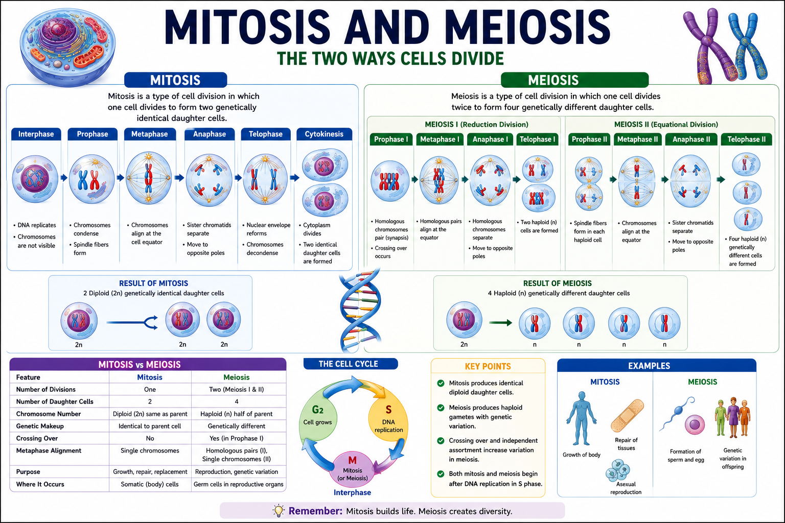
These two types of cell division are mitosis and meiosis, and together they represent the two fundamental ways that cells multiply. This mitosis and meiosis study guide is designed to give you a complete, clear, and genuinely useful understanding of both processes—every stage, every key event, every important difference—along with the biological context that makes the details memorable rather than just a list of phases to cram the night before an exam.
Here’s the honest truth about why students struggle with this topic: it’s not because mitosis and meiosis are inherently difficult. It’s because they’re usually taught as separate lists of phases, disconnected from any underlying logic. Once you understand why each division type exists—mitosis for growth and repair, meiosis for generating genetic diversity in sexual reproduction—and why each phase does what it does, everything clicks into place.
Throughout this guide, we’ll cover the complete cell cycle, walk through every stage of mitosis and meiosis with clear descriptions, explain crossing over and independent assortment, discuss what happens when cell division goes wrong, compare both processes side by side in a detailed table, and give you practice questions and a revision checklist to ensure exam readiness.
Whether you’re preparing for AP Biology, A-levels, MCAT, nursing boards, or a straightforward biology exam, this guide covers everything you need in one place. Let’s start at the very beginning—with what cell division actually is and why it matters so profoundly.
Key Takeaways
By the end of this guide, you’ll be able to:
- Explain why cell division is essential for growth, repair, and reproduction
- Describe all phases of the cell cycle including G1, S, G2, and M phases
- Walk through every stage of mitosis from prophase to cytokinesis with confidence
- Explain both divisions of meiosis (Meiosis I and II) and all their sub-stages
- Compare mitosis and meiosis across ten major features
- Explain crossing over and independent assortment and why they generate genetic variation
- Describe nondisjunction and the chromosomal disorders it causes
- Distinguish between sister chromatids, homologous chromosomes, diploid cells, and haploid cells
- Avoid the most common exam mistakes in cell division questions
What Is Cell Division?
Cell division is the biological process by which a parent cell divides into two or more daughter cells. It’s one of the most fundamental processes in all of biology—the mechanism by which life grows, replaces damaged cells, and reproduces.
All cells come from pre-existing cells—this is one of the core principles of cell theory (Virchow, 1855). Cell division is how pre-existing cells produce new ones. The process involves carefully copying the cell’s genetic material and then distributing those copies to daughter cells, ensuring each receives the information it needs to function.
There are two distinct types of cell division in eukaryotes:
- Mitosis – Produces two genetically identical daughter cells with the same chromosome number as the parent cell; used for growth, repair, and asexual reproduction
- Meiosis – Produces four genetically unique daughter cells with half the chromosome number of the parent cell; used exclusively for producing gametes (sex cells) in sexually reproducing organisms
Before either type of cell division can occur, the cell must duplicate its DNA—ensuring each daughter cell will receive a complete genome. This DNA replication happens during the S phase of the cell cycle, before the division phase begins.
Understanding the difference between these two division types—and why each exists—is the foundation for everything else in this guide.
Why Cell Division Is Important
Cell division isn’t just a biological curiosity—it’s the engine of life at the cellular level.
Growth and Development
Every multicellular organism begins as a single fertilized cell (zygote). Through repeated rounds of mitosis, that single cell becomes a trillion-cell organism. The selective activation of different genes in different cell lineages, combined with precise regulation of cell division, is what transforms a fertilized egg into a fully formed organism with hundreds of different cell types.
Tissue Repair and Regeneration
Skin heals after a cut because cells near the wound divide mitotically to replace what’s been lost. Bone fractures mend because osteoblast precursors divide and differentiate. Liver cells are remarkable—they can regenerate an entire liver from roughly 25% of the original tissue through mitosis.
Sexual Reproduction and Genetic Diversity
Meiosis produces gametes—sperm and eggs—that carry unique combinations of genetic material. Fertilization combines two haploid gametes into a diploid zygote with a completely new genetic combination. This genetic shuffling is the raw material of evolution and the reason sexually reproducing populations can adapt to changing environments.
Cell Replacement
Many cells have limited lifespans. Red blood cells live about 120 days; cells lining the gut are replaced every 3–5 days; white blood cells are continuously produced in bone marrow. Continuous mitosis maintains these cell populations.
History of Cell Division Discovery
The story of how scientists came to understand cell division spans over a century of increasingly sophisticated observation.
Walther Flemming (1882)
German anatomist Walther Flemming, working with salamander cells and aniline dyes, was the first to observe and document mitosis in detail. He described the behavior of chromosomes during division and coined the term mitosis (from the Greek mitos, meaning thread—referring to the thread-like appearance of chromosomes). His book Zellsubstanz, Kern und Zelltheilung (Cell Substance, Nucleus, and Cell Division) remains a landmark in cell biology.
Eduard van Beneden (1883)
Van Beneden observed meiosis in roundworm eggs (Ascaris megalocephala) and demonstrated that gametes contain half the chromosome number of somatic cells—the first observation of chromosome reduction during reproduction.
August Weismann (1890s)
Weismann theorized that sexual reproduction requires a reduction division (what we now call meiosis) to prevent chromosome number from doubling with each generation. He predicted the existence of meiosis before its details were fully worked out.
Theodor Boveri and Walter Sutton (1902–1903)
Independently, these two scientists proposed the chromosome theory of heredity—that chromosomes are the carriers of Mendel’s hereditary factors (genes). Their work directly connected cell division to genetics.
Calvin Bridges and others (early 1900s)
Work with Drosophila (fruit flies) demonstrated nondisjunction—the failure of chromosomes to separate properly during meiosis—and confirmed the chromosomal basis of heredity.
What Is Mitosis?
Mitosis is the type of cell division that produces two daughter cells genetically identical to the parent cell and to each other. It’s the primary mechanism of cell multiplication in somatic (non-reproductive) cells.
Purpose of Mitosis
Mitosis serves three main purposes:
- Growth – Increasing cell number during development
- Repair and replacement – Replacing dead or damaged cells in existing tissues
- Asexual reproduction – In unicellular organisms, mitosis IS reproduction; in some multicellular organisms, it enables vegetative reproduction (budding in Hydra, runner formation in strawberries)
Where Mitosis Occurs
In multicellular organisms, mitosis occurs primarily in:
- Stem cells of various tissues (bone marrow, intestinal crypts, skin basal layer)
- Embryonic cells during development
- Meristematic zones in plants (shoot apical meristems, root apical meristems)
Not all cells retain the ability to divide. Neurons in the adult brain, cardiac muscle cells, and mature red blood cells (which have no nucleus) are either permanently non-dividing or have very limited mitotic capacity.
Importance of Mitosis
- Maintains the diploid chromosome number (2n) in daughter cells
- Ensures genetic continuity—all cells in a multicellular organism carry identical DNA (mutations aside)
- Enables wound healing, tissue renewal, and organ development
Stages of Mitosis
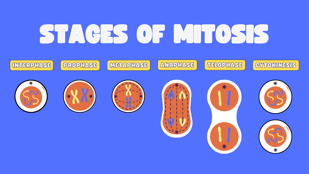
Before mitosis begins, the cell goes through interphase—a period of growth and preparation that’s often overlooked but absolutely critical.
Interphase
Interphase is not a stage of mitosis—it’s the period before mitosis. But no description of cell division is complete without it, because it’s where the cell spends most of its life and where all the preparation for division happens.
Interphase has three sub-phases:
- G₁ (Gap 1) – Cell grows; produces proteins and organelles; a checkpoint determines whether conditions are right for division
- S phase (Synthesis) – DNA is replicated; each chromosome is duplicated to form two identical sister chromatids joined at the centromere
- G₂ (Gap 2) – Cell continues growing; produces mitotic proteins; another checkpoint verifies DNA replication accuracy
By the end of interphase, the cell has doubled its DNA content and is ready to divide.
Prophase
Prophase is the first and longest stage of mitosis. Several dramatic changes occur:
- Chromatin condenses into visible, distinct chromosomes. Each chromosome now consists of two identical sister chromatids joined at the centromere.
- The mitotic spindle begins to form – In animal cells, centrioles duplicate and move to opposite poles; spindle fibers (microtubules) extend from the centrioles
- The nuclear envelope breaks down – This allows spindle fibers to access chromosomes
- The nucleolus disappears
Exam Tip: Prophase is when chromosomes first become visible under a microscope. If an exam shows a cell with condensed chromosomes but no alignment yet, it’s in prophase.
Metaphase
Metaphase is the most visually distinctive and most photographed stage of mitosis because chromosomes are at their most compact and most organized:
- Chromosomes line up along the cell’s equatorial plate (also called the metaphase plate)—an imaginary plane at the midpoint of the cell
- Spindle fibers (kinetochore fibers) attach to the centromeres of each chromosome at protein complexes called kinetochores
- Each chromosome is held in tension by fibers pulling from both poles
Metaphase is the ideal stage for karyotyping—creating a map of an organism’s chromosomes—because chromosomes are maximally condensed and clearly visible.
Anaphase
Anaphase is the stage that separates sister chromatids:
- Centromeres split – Sister chromatids are separated
- Former sister chromatids are pulled to opposite poles by shortening of spindle fibers
- Each pole now receives one complete set of chromosomes
- The cell elongates as spindle fibers push the poles apart
- At the end of anaphase, each pole has a complete, identical set of chromosomes
Important Fact: Anaphase is the stage where chromosomes (now individual chromatids) move toward the poles. If you see chromosomes at opposite poles of the cell but the cell hasn’t yet divided, it’s anaphase (or early telophase).
Telophase
Telophase is essentially the reverse of prophase:
- Chromosomes arrive at opposite poles and begin to decondense (uncoil)
- Nuclear envelopes reform around each set of chromosomes
- Nucleoli reappear in each forming nucleus
- The spindle apparatus breaks down
At the end of telophase, the cell has two nuclei—each containing a complete, identical set of chromosomes.
Cytokinesis
Cytokinesis is the division of the cytoplasm, separating the cell into two distinct daughter cells. It typically begins during late anaphase and completes after telophase.
In animal cells:
A contractile ring of actin and myosin filaments forms at the cell’s midpoint, pinching the cell membrane inward—creating a cleavage furrow that deepens until the cell is divided in two.
In plant cells:
The rigid cell wall prevents cleavage furrow formation. Instead, Golgi-derived vesicles fuse along the cell’s equatorial plate, depositing cell wall material to form a cell plate that develops into a new cell wall between the daughter cells.
Result of Mitosis
| Feature | Value |
|---|---|
| Number of daughter cells | 2 |
| Chromosome number | Same as parent (diploid → diploid, or haploid → haploid) |
| Genetic relationship to parent | Genetically identical (barring mutation) |
| Type of cells produced | Somatic cells |
| Function | Growth, repair, replacement, asexual reproduction |
What Is Meiosis?
Meiosis is the specialized type of cell division that produces four haploid daughter cells from a single diploid parent cell. These daughter cells are genetically unique—no two are alike—and they serve as gametes (eggs and sperm) in animals, or as spores in plants and fungi.
Purpose of Meiosis
Meiosis has two interrelated purposes:
- Reduce chromosome number from diploid (2n) to haploid (n)—so that when two gametes fuse at fertilization, the resulting zygote has the correct diploid number rather than doubling it each generation
- Generate genetic diversity—through crossing over and independent assortment—providing the variation that natural selection can act upon
Where Meiosis Occurs
In humans and other animals:
- Males: In the testes, during spermatogenesis (producing sperm)
- Females: In the ovaries, during oogenesis (producing eggs)
In plants:
- In sporangia (structures producing spores)—meiosis produces haploid spores, not gametes directly (gametes form later through mitosis of the haploid generation)
Importance of Meiosis
- Maintains the correct chromosome number across generations in sexually reproducing organisms
- Generates enormous genetic diversity—essential for adaptation and evolution
- Enables sexual reproduction—the dominant reproductive strategy of most complex multicellular life
Meiosis I Explained
Meiosis consists of two rounds of division—Meiosis I and Meiosis II—preceded by a single round of DNA replication during interphase.
Meiosis I is the reductional division—this is where the chromosome number is halved. Homologous chromosomes separate (rather than sister chromatids, as in mitosis).
Prophase I
Prophase I is the longest and most complex phase in all of meiosis. Several crucial events occur:
Chromosome condensation – Chromosomes condense, as in mitotic prophase.
Synapsis – Homologous chromosomes (pairs of chromosomes with the same genes, one from each parent) pair up along their entire length through a process called synapsis. The resulting structure of paired homologs is called a bivalent or tetrad (because it contains four chromatids—two from each homolog).
Crossing over (recombination) – While homologs are synapsed, chromatids from homologous chromosomes exchange segments of DNA at points called chiasmata (singular: chiasma). This creates recombinant chromosomes with new combinations of alleles—a major source of genetic variation.
Spindle formation – The nuclear envelope breaks down; spindle fibers form.
Exam Tip: Crossing over ONLY occurs during Prophase I of meiosis. Never during mitosis. If an exam asks when genetic recombination occurs, the answer is Prophase I.
Metaphase I
- Tetrads (bivalents) align at the metaphase plate—but unlike mitosis, it’s pairs of homologous chromosomes that align, not individual chromosomes
- Spindle fibers attach to the centromere of each homolog (one fiber from each pole to one chromosome of each homologous pair)
- Independent assortment determines how each pair of homologs aligns—which homolog faces which pole is random
This random orientation is called random independent assortment and is the second major source of genetic variation in meiosis.
Anaphase I
- Homologous chromosomes are pulled apart to opposite poles—not sister chromatids (as in mitosis)
- Sister chromatids remain joined at their centromeres and move together to the same pole
- Each pole receives ONE chromosome from each homologous pair
This is the key moment of chromosome reduction—the chromosome number is halved.
Telophase I
- Chromosomes arrive at opposite poles
- In many organisms, nuclear envelopes reform and the cell undergoes cytokinesis, producing two haploid cells (each still with replicated—double chromatid—chromosomes)
- In some organisms, telophase I is brief or skipped, and cells move directly into Meiosis II
After Meiosis I, there is a brief period called interkinesis—NO additional DNA replication occurs. This is crucial—it’s different from interphase before Meiosis I.
Meiosis II Explained
Meiosis II is essentially a mitotic division of the two haploid cells produced by Meiosis I. Its purpose is to separate sister chromatids.
Prophase II
- Chromosomes condense (if they decondensed after Meiosis I)
- Nuclear envelopes break down
- Spindle apparatus forms in each cell
Metaphase II
- Individual chromosomes (each consisting of two sister chromatids) align at the metaphase plate
- Spindle fibers attach to centromeres
- This looks similar to mitotic metaphase—except the cells are now haploid
Anaphase II
- Centromeres split; sister chromatids are pulled to opposite poles
- This is now identical in mechanism to mitotic anaphase
- Each pole receives one chromatid from each chromosome
Telophase II
- Chromatids arrive at poles and decondense
- Nuclear envelopes reform around each set of chromosomes
- Four haploid nuclei are now present across the two cells
Cytokinesis
- Both cells from Meiosis I divide
- Four haploid (n) daughter cells are produced
- Each cell contains one chromatid (now a single-stranded chromosome) from each homologous pair
Result of Meiosis
| Feature | Value |
|---|---|
| Number of daughter cells | 4 |
| Chromosome number | Half the parent cell (diploid → haploid) |
| Genetic relationship | Genetically unique—different from parent and each other |
| Type of cells produced | Gametes (animals) or spores (plants, fungi) |
| Function | Sexual reproduction; generating genetic diversity |
Mitosis vs Meiosis (Detailed Comparison Table)
| Feature | Mitosis | Meiosis |
|---|---|---|
| Number of divisions | 1 | 2 (Meiosis I + Meiosis II) |
| Number of daughter cells | 2 | 4 |
| Chromosome number in daughter cells | Same as parent (2n → 2n) | Half of parent (2n → n) |
| DNA replication | Once, before division | Once before Meiosis I; none before Meiosis II |
| Crossing over | Does not occur | Occurs during Prophase I |
| Genetic variation | Daughter cells are genetically identical | Daughter cells are genetically unique |
| Synapsis of homologs | Does not occur | Occurs during Prophase I |
| Alignment at metaphase plate | Individual chromosomes | Homologous pairs (Meiosis I) |
| What separates in anaphase | Sister chromatids | Homologs (Meiosis I); Sister chromatids (Meiosis II) |
| Cell type produced | Somatic (body) cells | Gametes or spores |
| Purpose | Growth, repair, asexual reproduction | Sexual reproduction; genetic diversity |
| Location in humans | All dividing somatic tissues | Gonads (testes, ovaries) |
| Cytokinesis | Once | Twice (after Meiosis I and II) |
| Interphase between divisions | N/A | Interkinesis (no DNA replication) |
| Result | Genetically identical clones | Genetically diverse haploid cells |
Similarities Between Mitosis and Meiosis
Despite their significant differences, mitosis and meiosis share several fundamental features:
- Both are preceded by DNA replication during S phase
- Both involve chromosome condensation at the start of the M phase
- Both use spindle fibers to move chromosomes
- Both involve nuclear envelope breakdown and reformation
- Both end with cytokinesis (though it occurs twice in meiosis)
- Both involve the same basic phases: prophase, metaphase, anaphase, telophase
- Both occur in eukaryotic cells (prokaryotes use binary fission)
- Both require ATP to power the molecular motors moving chromosomes
- Sister chromatids are separated in both (in mitotic anaphase and in Meiosis II anaphase)
Chromosome Behavior During Cell Division
Understanding how chromosomes behave requires clarity on some key terms that students frequently confuse.
Essential vocabulary:
- Chromosome – A single piece of DNA with associated proteins; humans have 46 chromosomes (23 pairs)
- Chromatid – One half of a replicated chromosome; two chromatids joined at the centromere make one replicated chromosome
- Sister chromatids – The two identical chromatids formed by DNA replication of a single chromosome; they’re joined at the centromere
- Homologous chromosomes – Pairs of chromosomes that carry the same genes (one from each parent); they’re similar but not identical (may have different alleles)
- Diploid (2n) – Having two copies of each chromosome; human somatic cells are 2n=46
- Haploid (n) – Having one copy of each chromosome; human gametes are n=23
- Centromere – The region where sister chromatids are attached and where spindle fibers connect
- Kinetochore – Protein complex at the centromere where spindle fibers attach
- Bivalent/Tetrad – A pair of synapsed homologous chromosomes during Meiosis I, consisting of four chromatids total
Chromosome number changes:
In mitosis: 2n → 2n (46 → 46 in humans)
In Meiosis I: 2n → n (46 → 23)
In Meiosis II: n → n (23 → 23, but sister chromatids are now separated)
Crossing Over Explained
Crossing over (genetic recombination) is one of the most important events in sexual reproduction. It occurs during Prophase I of meiosis, when homologous chromosomes are synapsed (paired together).
How crossing over works:
- Homologous chromosomes pair up during synapsis in Prophase I, forming a tetrad (bivalent) of four chromatids
- Non-sister chromatids from homologous chromosomes physically exchange segments at points called chiasmata (singular: chiasma)
- The exchanged segments are complementary—the same genes, possibly with different alleles
- The resulting chromosomes are recombinant chromosomes—they contain allele combinations that didn’t exist in either parent chromosome
Why crossing over matters:
- Creates new combinations of alleles on chromosomes—recombinant genotypes
- A single chromosome pair typically undergoes 1–3 crossovers per meiosis
- With 23 pairs of chromosomes in humans, crossing over alone can produce an astronomically large number of unique gametes
- Crossing over is used by geneticists to map gene locations—the further apart two genes are on a chromosome, the more likely crossing over will separate them (and the higher the recombination frequency)
Practical example: If one chromosome carries alleles for brown hair and blue eyes, and its homolog carries alleles for blonde hair and brown eyes, crossing over can produce chromosomes carrying brown hair + brown eyes or blonde hair + blue eyes—combinations that didn’t exist in either parental chromosome.
Independent Assortment Explained
Independent assortment is the second major mechanism generating genetic diversity during meiosis. It operates during Metaphase I.
How independent assortment works:
When homologous pairs line up at the metaphase plate during Meiosis I, the orientation of each pair is completely random. Either chromosome of a homologous pair can face either pole—independently of every other homologous pair.
The mathematics of independent assortment:
- For each homologous pair, there are 2 possible orientations
- For n pairs of chromosomes, there are 2ⁿ possible combinations
- In humans, with 23 pairs: 2²³ = approximately 8.4 million different possible gametes from independent assortment alone
When combined with crossing over (which occurs simultaneously), the number of genetically distinct gametes a human can produce is essentially limitless—far greater than 8.4 million. This is why no two humans (except identical twins) are genetically identical.
Mendel’s Law of Independent Assortment describes this phenomenon at the gene level: alleles of different genes assort independently during gamete formation—provided the genes are on different chromosomes (or far apart on the same chromosome).
Importance of Mitosis in Living Organisms
Development: Every multicellular organism develops through repeated rounds of mitosis starting from a single fertilized egg. The human body contains approximately 37 trillion cells—all derived from mitotic divisions of that original zygote.
Wound healing: When you cut yourself, cells at the wound margins are stimulated to divide mitotically, filling in the gap. Growth factors released by platelets and damaged cells trigger nearby cells to re-enter the cell cycle.
Organ regeneration: The liver has remarkable regenerative capacity—if up to 70% is removed, the remaining tissue can regenerate fully within weeks through mitosis. This makes living-donor liver transplantation possible.
Immune response: When your immune system detects a pathogen, specific immune cells (lymphocytes that recognize that pathogen) undergo rapid mitosis—clonal expansion—producing armies of cells to fight the infection.
Asexual reproduction: Organisms like bacteria (binary fission—equivalent to mitosis in prokaryotes), yeast (budding), and many plants (vegetative reproduction) use mitosis as their primary or sole mode of reproduction.
Importance of Meiosis in Sexual Reproduction
Chromosome number maintenance: Without meiosis, chromosome numbers would double with every generation of sexual reproduction. Meiosis halves the chromosome number in gametes; fertilization restores the diploid number—maintaining genomic stability across generations.
Genetic diversity: Every sexually reproducing organism that has ever lived is genetically unique (barring identical twins and other identical clonal organisms). This diversity means populations can adapt to changing environments—natural selection can favor individuals with advantageous combinations.
Evolution: Genetic variation generated by meiosis is the raw material of evolution. Without recombination and independent assortment, all individuals in a sexually reproducing species would carry essentially the same genes.
Disease resistance: Genetic diversity means no pathogen can eliminate an entire sexually reproducing population—some individuals will always have combinations of alleles conferring partial resistance.
Cell Cycle and Cell Division
The cell cycle is the ordered sequence of events that a dividing cell goes through from one division to the next. Understanding it provides context for both mitosis and meiosis.
G1 Phase (Gap 1)
- Cell grows and increases in size
- Synthesizes proteins and RNA
- Organelles are duplicated
- G1 checkpoint (restriction point): The cell assesses whether it has adequate nutrients, growth signals, and undamaged DNA before committing to division. This is the main regulatory checkpoint—most cells that will divide commit here; cells that won’t divide enter G₀.
S Phase (DNA Synthesis)
- DNA replication occurs—each chromosome is duplicated to produce two identical sister chromatids
- Histone proteins are also synthesized
- Duration: approximately 8 hours in human cells
- No other checkpoint—once S phase begins, the cell is committed to completing division
G2 Phase (Gap 2)
- Cell continues to grow
- Produces proteins needed for mitosis (e.g., tubulin for spindle fibers)
- G2 checkpoint: Verifies that DNA replication is complete and accurate; checks for DNA damage; if damage is detected, p53 (a tumor suppressor protein) can halt the cycle or trigger apoptosis
M Phase (Mitotic Phase)
- Encompasses mitosis (PMAT) and cytokinesis
- Spindle assembly checkpoint (SAC): During metaphase, ensures all kinetochores are properly attached to spindle fibers before anaphase proceeds; prevents premature chromosome separation
Cell cycle regulation failures:
When checkpoints fail—due to mutations in genes like p53, RB (retinoblastoma protein), or BRCA1/2—cells can divide uncontrollably. This is the molecular basis of cancer. Understanding the cell cycle is therefore directly relevant to oncology.
Common Errors During Cell Division
Nondisjunction
Nondisjunction is the failure of chromosomes or chromatids to separate properly during cell division.
Nondisjunction can occur:
- During Meiosis I: Homologous chromosomes fail to separate → one gamete gets both chromosomes of a pair; the other gets none
- During Meiosis II: Sister chromatids fail to separate → similar imbalance
- During Mitosis: Sister chromatids fail to separate → produces daughter cells with incorrect chromosome numbers (aneuploidy) in somatic cells
Consequences of nondisjunction:
| Condition | Cause | Chromosome Number | Features |
|---|---|---|---|
| Down syndrome (Trisomy 21) | Extra chromosome 21 | 2n = 47 | Intellectual disability, characteristic facial features, heart defects |
| Edwards syndrome (Trisomy 18) | Extra chromosome 18 | 2n = 47 | Severe developmental defects; usually fatal in first year |
| Patau syndrome (Trisomy 13) | Extra chromosome 13 | 2n = 47 | Severe brain, heart, and other organ defects |
| Turner syndrome (45, X) | Missing X chromosome | 2n = 45 | Female; short stature, infertility, heart defects |
| Klinefelter syndrome (47, XXY) | Extra X chromosome in males | 2n = 47 | Male; reduced fertility, mild learning difficulties |
Incidence: Nondisjunction becomes more common as maternal age increases. This is why the risk of trisomy 21 increases with advanced maternal age—egg cells are arrested in Prophase I from fetal development until ovulation, and longer arrest times increase the risk of chromosome separation errors.
Mutation
DNA mutations can occur during replication if errors aren’t corrected by proofreading mechanisms or DNA repair systems. Replication errors, chemical mutagens, radiation, and other factors can introduce mutations that affect gene function, including genes that regulate the cell cycle itself.
Chromosomal Disorders
Beyond nondisjunction, chromosomal disorders can arise from structural abnormalities:
- Deletions – Loss of a chromosomal segment
- Duplications – Extra copy of a chromosomal segment
- Inversions – A segment is reversed within a chromosome
- Translocations – A segment moves to a non-homologous chromosome
Example: The Philadelphia chromosome in chronic myelogenous leukemia (CML) results from a translocation between chromosomes 9 and 22, creating a fusion gene (BCR-ABL) encoding an overactive kinase that drives uncontrolled cell proliferation.
Mitosis and Meiosis in Humans
Mitosis in humans:
Occurs in all dividing somatic cells throughout life. Key examples:
- Skin cells: The basal layer of the epidermis divides constantly; cells produced migrate upward, differentiate, and are shed within weeks
- Bone marrow: Produces ~200 billion red blood cells per day through mitosis and differentiation
- Intestinal epithelium: Completely replaced every 3–5 days—one of the fastest cell turnover rates in the body
- Embryonic development: From fertilized egg to fully formed fetus through ~44 rounds of mitosis
Meiosis in humans:
Occurs only in gonads. The timing differs significantly between sexes:
- Males (spermatogenesis): Meiosis begins at puberty and continues throughout life. Each primary spermatocyte (2n) produces four functional sperm (n). The entire process takes approximately 74 days. Males produce ~300 million sperm per day.
- Females (oogenesis): A striking difference—meiosis begins during fetal development (around 3–5 months gestation), but oocytes are arrested in Prophase I until after puberty. At ovulation, each month one primary oocyte completes Meiosis I (producing a secondary oocyte and a polar body) and begins Meiosis II—but is arrested again in Metaphase II. Meiosis II is only completed if fertilization occurs. Of the original ~7 million oocytes in a female fetus, only ~400–500 will ever ovulate; the rest undergo atresia.
Mitosis and Meiosis in Plants
Plants have a unique life cycle called alternation of generations, which means mitosis and meiosis play different roles than in animals.
Mitosis in plants:
- Occurs in meristematic tissues: shoot apical meristems (tip of shoots), root apical meristems, lateral meristems (cambium—for secondary growth in woody plants)
- Unlike animals, plants grow throughout their lives through localized mitosis at meristems
- Also occurs in vegetative reproduction (runners, bulbs, rhizomes)
Meiosis in plants:
- Occurs in sporangia (spore-producing structures in reproductive organs)
- Produces haploid spores (not gametes directly)
- Spores grow by mitosis into the gametophyte generation—a haploid multicellular plant
- Gametes are produced by the gametophyte through mitosis
- Fertilization of gametes produces the diploid sporophyte generation
In flowering plants:
- Meiosis in the anther produces microspores (male, which develop into pollen grains)
- Meiosis in the ovule produces megaspores (female, which develop into the embryo sac containing the egg cell)
Common Biology Terms Every Student Should Know
| Term | Definition |
|---|---|
| Chromatin | Loosely packed DNA associated with proteins; found in interphase nucleus |
| Chromosome | Condensed, organized structure of DNA and protein; visible during cell division |
| Sister chromatids | Two identical chromatids joined at centromere; produced by DNA replication |
| Homologous chromosomes | Chromosome pairs carrying the same genes (but possibly different alleles); one from each parent |
| Diploid (2n) | Cell containing two sets of chromosomes |
| Haploid (n) | Cell containing one set of chromosomes |
| Centromere | Region joining sister chromatids; point of spindle fiber attachment |
| Kinetochore | Protein complex at centromere; spindle fiber binding site |
| Spindle fibers | Microtubule structures that move chromosomes during cell division |
| Synapsis | Pairing of homologous chromosomes during Prophase I of meiosis |
| Bivalent/Tetrad | Paired homologous chromosomes (4 chromatids total) during Meiosis I |
| Chiasma | Point of crossover between non-sister chromatids during Prophase I |
| Crossing over | Exchange of segments between non-sister chromatids of homologs |
| Independent assortment | Random orientation of homologous pairs at Metaphase I |
| Nondisjunction | Failure of chromosomes to separate properly during division |
| Aneuploidy | Having an abnormal number of chromosomes |
| Gamete | Haploid reproductive cell (sperm or egg) |
| Karyotype | Organized image of an organism’s chromosomes |
| Cell plate | Structure forming new cell wall in dividing plant cells |
| Cleavage furrow | Indentation in animal cell membrane during cytokinesis |
Common Mistakes Students Make
Here are the errors that consistently cost biology students marks in cell division questions.
1. Confusing homologous chromosomes with sister chromatids
Sister chromatids are the two identical copies of ONE chromosome produced by DNA replication—joined at the centromere. Homologous chromosomes are pairs of chromosomes that carry the same genes but came from different parents (one maternal, one paternal)—they may carry different alleles. In mitosis anaphase, sister chromatids separate. In Meiosis I anaphase, homologs separate. Get these mixed up and your entire understanding of cell division falls apart.
2. Saying crossing over occurs in mitosis
Crossing over occurs ONLY during Prophase I of meiosis. Never in mitosis. Mitotic chromosomes condense and align individually at the metaphase plate—they do not synapse with homologs, so crossing over cannot occur. This distinction is tested frequently.
3. Forgetting that Meiosis II is NOT preceded by DNA replication
After Meiosis I, cells enter interkinesis—there is no DNA replication. The purpose of Meiosis II is to separate the sister chromatids that are still joined from the original replication before Meiosis I. Students sometimes incorrectly state that DNA is replicated before both Meiosis I and II.
4. Stating that meiosis produces two cells
Meiosis produces four haploid cells (though in female animals, three of these become non-functional polar bodies, leaving only one functional egg). The confusion often comes from conflating Meiosis I (which does produce two cells) with the complete meiotic process.
5. Confusing chromosome number with chromatid number
A chromosome can be either single (one DNA molecule) or replicated (two sister chromatids joined at centromere). After S phase but before anaphase, a human cell has 46 chromosomes but 92 chromatids. At the end of mitotic anaphase, the cell has 92 chromosomes (now individual, single-chromatid chromosomes) before cytokinesis splits them into two sets of 46.
6. Incorrect PMAT sequence for meiosis
Students sometimes apply the simple PMAT mnemonic to meiosis and forget that it has two rounds: Meiosis I (PMAT-I) and Meiosis II (PMAT-II). Always specify which round when describing meiotic phases.
Best Tips to Study Mitosis and Meiosis
1. Learn the vocabulary first—everything else depends on it
Before you try to understand the stages, make sure you can define and distinguish: chromosome, chromatid, sister chromatid, homologous chromosome, centromere, kinetochore, diploid, haploid. Attempting to learn the stages without these terms is like trying to follow directions without knowing what a road is.
2. Draw every stage from memory
For each phase of mitosis and meiosis, draw a cell with a simple set of chromosomes (2 pairs is usually enough) and show what’s happening to the chromosomes. Doing this for all 4+8 stages is the single most effective way to cement this material. You can’t draw it accurately if you don’t understand it.
3. Use color coding
When drawing chromosome diagrams, use one color for maternal chromosomes and another for paternal chromosomes. This makes it immediately obvious what crossing over and independent assortment actually do to chromosome composition.
4. Build the comparison table yourself
Read through this guide, then close it and construct the mitosis vs meiosis comparison table from memory. Every feature you can’t recall points you to exactly what needs more review.
5. Connect nondisjunction to real clinical conditions
Don’t just know that nondisjunction produces aneuploidy—know that trisomy 21 causes Down syndrome, that Turner syndrome (45, X) is monosomy of the sex chromosome, that Klinefelter syndrome (47, XXY) involves an extra X. These clinical connections are tested frequently and make the abstract concept of nondisjunction tangible.
6. Practice chromosome number problems
Given a species with 2n=8, how many chromosomes are in each cell after Meiosis I? After Meiosis II? After mitosis? How many chromatids are present in a cell in G2? These calculations become straightforward once the underlying concepts are solid, but they require practice to execute quickly and accurately under exam pressure.
Mitosis and Meiosis Practice Questions
20 Multiple Choice Questions with Answers
- Which phase of mitosis do chromosomes align at the metaphase plate?
- A) Prophase
- B) Anaphase
- C) Metaphase ✓
- D) Telophase
- Crossing over occurs during which phase of meiosis?
- A) Metaphase I
- B) Prophase I ✓
- C) Anaphase II
- D) Prophase II
- How many haploid daughter cells result from one complete meiotic division?
- A) 2
- B) 3
- C) 4 ✓
- D) 8
- Which of the following correctly describes sister chromatids?
- A) Two homologous chromosomes paired during meiosis
- B) Two identical chromatids joined at a centromere ✓
- C) Two chromosomes from different parents
- D) Chromosomes from two different cell types
- What is the primary purpose of meiosis?
- A) Growth and tissue repair
- B) Asexual reproduction
- C) Production of genetically identical cells
- D) Production of genetically diverse haploid gametes ✓
- During which phase of mitosis does the nuclear envelope break down?
- A) Interphase
- B) Prophase ✓
- C) Metaphase
- D) Anaphase
- In which phase of Meiosis I are homologous chromosomes separated?
- A) Prophase I
- B) Metaphase I
- C) Anaphase I ✓
- D) Telophase I
- A human cell in G2 has how many chromosomes?
- A) 23
- B) 46 ✓
- C) 92
- D) 48
- Which of the following structures forms during cytokinesis in plant cells?
- A) Cleavage furrow
- B) Cell plate ✓
- C) Spindle fiber
- D) Centrosome
- Nondisjunction during Meiosis I results in:
- A) All gametes having the correct chromosome number
- B) Two gametes with an extra chromosome; two with a missing chromosome ✓
- C) Four gametes with an extra chromosome
- D) No effect on chromosome number
- Which of the following is a result of independent assortment?
- A) Identical daughter cells from mitosis
- B) Random distribution of homologs to gametes ✓
- C) Formation of sister chromatids
- D) DNA replication before cell division
- Down syndrome (Trisomy 21) results from:
- A) Deletion of chromosome 21
- B) Nondisjunction resulting in an extra chromosome 21 ✓
- C) Translocation of chromosome 14
- D) Inversion within chromosome 21
- What is the ploidy of cells produced at the end of Meiosis I?
- A) Diploid with sister chromatids
- B) Haploid with sister chromatids ✓
- C) Haploid without sister chromatids
- D) Diploid without sister chromatids
- Which cell cycle checkpoint verifies proper spindle attachment to chromosomes?
- A) G1 checkpoint
- B) G2 checkpoint
- C) Spindle assembly checkpoint ✓
- D) S phase checkpoint
- During mitotic anaphase, what separates and moves to opposite poles?
- A) Homologous chromosomes
- B) Sister chromatids ✓
- C) Bivalents
- D) Chiasmata
- The number of possible gamete combinations from independent assortment in humans is:
- A) 46
- B) 23
- C) 8.4 million ✓
- D) 2 million
- Which phase is unique to meiosis but does NOT occur in mitosis?
- A) Metaphase
- B) Anaphase
- C) Prophase I with synapsis ✓
- D) Telophase
- After DNA replication in S phase, each chromosome consists of:
- A) One chromatid
- B) Two sister chromatids ✓
- C) Two homologous chromosomes
- D) Four chromatids
- Turner syndrome (45, X) is caused by:
- A) An extra X chromosome in females
- B) A missing sex chromosome in females ✓
- C) An extra autosome
- D) A chromosomal translocation
- Which of the following processes generates the MOST genetic diversity?
- A) Mitosis
- B) DNA replication
- C) Cytokinesis
- D) Meiosis with crossing over and independent assortment ✓
10 Short Answer Questions
- Distinguish between sister chromatids and homologous chromosomes. Explain when each pair is separated during cell division.
- Explain why crossing over increases genetic variation. At what stage of meiosis does it occur, and what is the structural evidence (chiasma) for this event?
- A species has a diploid chromosome number of 2n=12. How many chromosomes are present in: (a) a cell in G1; (b) a cell in G2; (c) a secondary oocyte; (d) a mature sperm cell?
- Describe the spindle assembly checkpoint. What happens if this checkpoint fails, and what is the clinical consequence?
- Compare cytokinesis in plant and animal cells. Explain why the mechanisms differ and describe the structures involved in each.
- Explain why Meiosis I is called the “reductional division” while Meiosis II is called the “equational division.”
- What is nondisjunction? Describe how nondisjunction in Meiosis I differs from nondisjunction in Meiosis II in terms of the gametes produced.
- Explain how independent assortment and crossing over work together to generate virtually limitless genetic diversity in human gametes.
- Describe the role of the G1 checkpoint in preventing uncontrolled cell division. What molecular players enforce this checkpoint?
- Compare the products of spermatogenesis and oogenesis. How many functional cells result from each, and why is this difference adaptive?
5 Long Answer Questions
- Describe the complete process of mitosis from interphase through cytokinesis. For each phase, describe the behavior of chromosomes, spindle fibers, and the nuclear envelope. Explain what occurs during cytokinesis in both animal and plant cells. Discuss the significance of cell cycle checkpoints and explain what happens when they fail, using cancer as an example.
- Describe Meiosis I and Meiosis II in complete detail, covering all phases of each division. For each phase, explain the key events occurring with chromosomes, spindle fibers, and the nuclear envelope. Explain why DNA replication does NOT occur between Meiosis I and Meiosis II, and how this relates to the purpose of the two divisions.
- Compare mitosis and meiosis comprehensively, addressing the purpose, location, number of divisions, chromosome number in daughter cells, genetic outcome, DNA replication, crossing over, and synapsis. Explain why both processes are essential for life and what would happen to a population if meiosis produced diploid gametes instead of haploid gametes.
- Explain how genetic diversity is generated during meiosis. Describe crossing over (including the molecular events, the structures formed, and the significance of chiasmata), independent assortment (including the mathematical basis), and fertilization as a third source of variation. Calculate the theoretical number of genetically unique gametes a human can produce through independent assortment alone, and explain why the actual number is far greater.
- Discuss errors in cell division and their consequences. Describe nondisjunction during Meiosis I and Meiosis II—explaining how each produces gametes with abnormal chromosome numbers. Discuss three specific chromosomal disorders (include their chromosomal basis, common phenotypic features, and the role of maternal age where relevant). Also describe structural chromosomal mutations (deletion, duplication, inversion, translocation) and give one clinical example of each.
Revision Checklist
Use this checklist honestly before any exam on cell division.
- I can explain the purpose of mitosis and meiosis and when each is used
- I can describe the cell cycle (G1, S, G2, M) and explain what happens at each checkpoint
- I can draw and label all phases of mitosis from prophase to cytokinesis
- I can describe cytokinesis in both plant and animal cells
- I can draw and label all phases of Meiosis I and Meiosis II
- I can explain what happens during Prophase I that doesn’t happen in any other phase
- I can distinguish sister chromatids from homologous chromosomes
- I can explain crossing over: when it occurs, what structures are involved, and its significance
- I can explain independent assortment and calculate the number of possible gamete combinations
- I can compare mitosis and meiosis across at least ten features
- I can describe nondisjunction and name three chromosomal disorders it causes
- I can explain the difference in meiosis between males (spermatogenesis) and females (oogenesis)
- I can describe how mitosis differs in plants vs animals (meristems, cell plate vs cleavage furrow)
- I can define all 20 key terms from the glossary section
- I have completed at least 20 MCQs and 3 long answer questions from this guide
Best Books for Learning Cell Division
- “Molecular Biology of the Cell” by Alberts, Johnson, Lewis et al. – The comprehensive gold standard reference. Its chapters on the cell cycle, mitosis, and meiosis are exceptionally clear and backed by stunning electron micrographs and diagrams. Available free through NCBI Bookshelf online.
- Campbell Biology (any recent edition) – The definitive AP Biology and introductory college biology textbook. The chapters on cell division are well-structured, include excellent diagrams of each phase, and connect cell division to genetics and cancer biology.
- “The Cell: A Molecular Approach” by Cooper and Hausman – Strong on the molecular mechanisms behind cell division—spindle assembly, checkpoint proteins, cyclin-CDK regulation. Excellent for students who want deeper mechanistic understanding.
- “Genetics: From Genes to Genomes” by Hartwell et al. – Outstanding coverage of meiosis in the context of genetics and inheritance. Particularly strong on connecting meiotic events to Mendel’s laws, mapping genes, and chromosomal disorders.
- “Cell Biology” by Pollard and Earnshaw – Comprehensive coverage of the cytoskeleton and cell division machinery at a level suited for advanced undergraduates and graduate students. Exceptional for understanding the molecular motors involved in chromosome movement.
Free Online Biology Resources
- OpenStax Biology 2e – Cell Division Chapters – Free, peer-reviewed, comprehensive coverage of the cell cycle, mitosis, and meiosis. Includes figures, review questions, and critical thinking exercises.
- Khan Academy – Cell Division – Excellent video lessons covering the cell cycle, mitosis, and meiosis with integrated practice questions. Particularly strong for visual learners and AP Biology preparation.
- Biology LibreTexts – Cell Division – Open-access academic content covering cell division at introductory through advanced levels.
- HHMI BioInteractive – Meiosis Resources – World-class animations of cell division, including an exceptional meiosis animation that shows chromosome behavior in genuine three-dimensional detail. These are the best free visual resources available for this topic.
- NCBI Bookshelf – Molecular Biology of the Cell – Free access to one of the most authoritative cell biology texts. The chapters on the cell cycle, mitosis, and meiosis provide both mechanistic detail and clinical connections.
Frequently Asked Questions
1. What is the main difference between mitosis and meiosis?
Mitosis produces two genetically identical diploid daughter cells for growth and repair. Meiosis produces four genetically unique haploid daughter cells for sexual reproduction. Mitosis involves one division; meiosis involves two. Crossing over and independent assortment occur in meiosis but not mitosis.
2. How many chromosomes do human cells have after mitosis vs meiosis?
After mitosis: 46 chromosomes (diploid—same as parent). After meiosis: 23 chromosomes (haploid—half the parent number). Fertilization of two haploid gametes restores the diploid number.
3. Does crossing over occur in mitosis?
No. Crossing over occurs exclusively during Prophase I of meiosis, when homologous chromosomes are synapsed. Mitotic chromosomes do not synapse with their homologs, so crossing over cannot occur.
4. What is the purpose of meiosis?
Meiosis serves two purposes: (1) it reduces chromosome number from diploid to haploid so that fertilization produces the correct diploid number; and (2) it generates genetic diversity through crossing over and independent assortment, providing the variation that drives evolution.
5. What causes Down syndrome?
Down syndrome (Trisomy 21) is caused by nondisjunction—the failure of chromosome 21 to separate properly during meiosis, most commonly Meiosis I in the egg. The resulting gamete carries two copies of chromosome 21; when fertilized by a normal sperm, the zygote has three copies (2n=47). Risk increases with maternal age.
6. Why do females have polar bodies in oogenesis?
The unequal cytoplasmic division during oogenesis produces one large secondary oocyte (which becomes the egg) and small, non-functional polar bodies that are eventually degraded. The unequal division concentrates the cytoplasm, nutrients, and organelles in one cell—maximizing the egg’s ability to support early embryonic development.
7. What is the difference between interphase and interkinesis?
Interphase is the period before meiosis I (and before mitosis), during which DNA replication (S phase) occurs. Interkinesis is the brief period between Meiosis I and Meiosis II—during which there is NO DNA replication. This distinction is crucial and frequently tested.
8. What happens at the G1 checkpoint?
The G1 checkpoint (restriction point) assesses whether the cell has adequate nutrients, growth factors, and undamaged DNA before committing to DNA replication and division. If conditions are unfavorable, the cell halts. In cancer, this checkpoint is often inactivated, allowing cells to divide regardless of damage or growth signals.
9. How many genetically distinct gametes can a human produce?
From independent assortment alone: 2²³ = ~8.4 million possible combinations. When crossing over is also considered, the number of genetically distinct gametes a human could theoretically produce is effectively infinite—far greater than 8.4 million. This explains why every person (except identical twins) is genetically unique.
10. What is Turner syndrome and how does it arise?
Turner syndrome (45, X) occurs when a female has only one X chromosome instead of two. It arises from nondisjunction during meiosis in either parent—the gamete that contributes to the affected individual had no sex chromosome. Characteristics include short stature, absent or underdeveloped ovaries, infertility, and sometimes heart defects.
11. Why is the spindle assembly checkpoint important?
The spindle assembly checkpoint ensures that all kinetochores on chromosomes are properly attached to spindle fibers before anaphase proceeds. If chromosomes are not properly attached, the checkpoint halts division. Without this checkpoint, chromosomes could be distributed unequally between daughter cells—producing aneuploid cells, which can contribute to cancer and other disorders.
12. Can mitosis produce haploid cells?
Yes—mitosis is not inherently diploid-specific. If a haploid cell divides by mitosis, it produces haploid daughter cells. This happens in plant gametophytes (the haploid phase of the plant life cycle), which grow by mitosis. In animals, all gametes are produced by meiosis—but if a haploid gamete were to divide, it would do so by mitosis.
Summary
This complete mitosis and meiosis study guide has covered the full landscape of cell division—from the cell cycle that prepares cells for division through every stage of both processes, the mechanisms generating genetic diversity, the consequences of errors, and the clinical significance of what happens when division goes wrong.
The essential distinction to carry forward: mitosis produces two genetically identical diploid cells for growth, repair, and replacement. Meiosis produces four genetically unique haploid cells for sexual reproduction. Meiosis involves two divisions (I and II), with crossing over during Prophase I and independent assortment during Metaphase I generating diversity that makes every sexually reproducing individual genetically unique.
The cell cycle (G1 → S → G2 → M) governs when and how cells divide, with checkpoints ensuring accuracy. When checkpoints fail—as they do in cancer—cell division becomes uncontrolled. Nondisjunction during meiosis produces aneuploid gametes, leading to conditions like Down syndrome, Turner syndrome, and Klinefelter syndrome.
Mitosis and meiosis are not just exam topics. They’re the molecular mechanisms of life, growth, reproduction, and heredity—the processes that have generated every living thing on Earth and every unique individual among them.
Final Thoughts
Cell division is where genetics becomes physical—where DNA sequences become chromosomes, chromosomes become daughter cells, and daughter cells become organisms. Mitosis and meiosis together account for everything from how a wound heals to how a new life begins to why siblings look similar but not identical.
Students who invest time in genuinely understanding both processes—not just memorizing the phases but understanding why each event happens and what it accomplishes—consistently find that the rest of genetics, embryology, and medicine falls into place more easily. Everything connects to cell division.
Use the revision checklist. Work through every practice question, especially the long answers that require you to explain your reasoning. Draw the phases until they become second nature. And connect every abstract concept—crossing over, independent assortment, nondisjunction—to something concrete: a genetic disorder, a forensic application, a disease treatment.
Understanding how cells divide is understanding the most fundamental machinery of life. It’s worth the effort.
Good luck with your studies.
References
- OpenStax Biology 2e – Cell Division – openstax.org/books/biology-2e
- Khan Academy – Cell Division – khanacademy.org
- Biology LibreTexts – Cell Division – bio.libretexts.org
- HHMI BioInteractive – Cell Division Resources – biointeractive.org
- NCBI Bookshelf – Molecular Biology of the Cell – ncbi.nlm.nih.gov/books
Article Disclaimer
This article is intended for educational and informational purposes only. While LearnMinto strives to provide accurate, well-researched, and up-to-date information, cell biology and genetics are continually refined through ongoing scientific research. The content is designed to support learning, revision, and exam preparation and should not replace official textbooks, classroom instruction, laboratory training, or guidance from qualified educators. Readers are encouraged to verify important academic concepts through trusted educational resources, recognized examination boards, and reputable scientific organizations before relying on this content for academic purposes. LearnMinto is not affiliated with any specific school, university, research institution, or examination board. This article is intended solely for educational purposes and should not be considered professional scientific, medical, or academic advice.