
Introduction
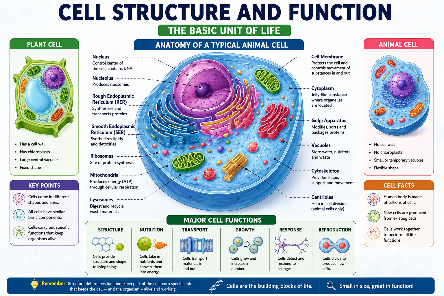
Every living thing you’ve ever seen—a soaring eagle, a blade of grass pushing through concrete, a bacterium invisible to the naked eye, and yes, you reading this right now—is built from cells. The cell is the fundamental unit of life, and understanding cell structure and function is essentially understanding the basic grammar of all biology.
Here’s the thing though: most students encounter cells for the first time through a textbook diagram that makes the whole thing look deceptively simple. A blob with some labeled dots inside. How hard can it be? Then the exam arrives, and suddenly the differences between rough and smooth endoplasmic reticulum matter enormously, and nobody can remember whether it’s xylem or phloem that— wait, wrong topic. The point is, cells are more complex and more fascinating than they first appear, and learning them properly the first time saves a lot of pain later.
This guide is built to fix that. Whether you’re studying for a high school biology exam, preparing for a medical entrance test, or trying to nail a college cell biology course, this is the one resource that should get you there without needing to open five other tabs.
We’ll cover everything: the history of cell theory, the structure of prokaryotic and eukaryotic cells, every major organelle and what it actually does, how materials move in and out of cells, how cells divide, how they produce energy, and much more. Along the way, you’ll find comparison tables, real-life examples, practice questions, and a revision checklist you can use right before any exam.
One cell at a time—let’s get started.
Key Takeaways
By the end of this guide, you’ll be able to:
- Explain cell theory and the scientists who developed it
- Distinguish between prokaryotic and eukaryotic cells with confidence
- Identify and describe the function of every major cell organelle
- Compare plant and animal cells accurately
- Explain six mechanisms of cell transport across the membrane
- Describe the cell cycle including interphase, mitosis, and cytokinesis
- Understand how cells produce energy through cellular respiration
- Define cell specialization and explain its importance in multicellular organisms
- Avoid the most common mistakes students make in cell biology exams
What Is a Cell?
A cell is the smallest structural and functional unit of all living organisms. Every biological process that keeps you alive—breathing, digesting food, thinking, healing from a cut—involves cells performing specific tasks with remarkable precision.
The word “cell” comes from the Latin cella, meaning small room. That name was coined by Robert Hooke in 1665 when he looked at a thin slice of cork through a simple microscope and saw tiny compartments that reminded him of the small rooms where monks lived in monasteries. He couldn’t have known at the time just how profound his observation was.
Some organisms—bacteria, for example—consist of just one cell. That single cell manages to reproduce, respond to the environment, acquire nutrients, and do everything else necessary to survive. At the other extreme, the human body contains approximately 37 trillion cells, each specialized for specific functions, communicating and cooperating in extraordinarily complex ways.
Cells come in a staggering variety of shapes and sizes, all dictated by their function:
- Red blood cells are biconcave discs—their flattened shape maximizes surface area for oxygen exchange and allows them to flex through narrow capillaries
- Neurons have long, branching extensions (axons and dendrites) that allow them to transmit electrical signals across long distances
- Muscle cells are elongated with abundant mitochondria to power contraction
- Root hair cells have long projections that increase surface area for water absorption
Shape follows function in biology, and cells are the clearest demonstration of that principle.
Cell Theory
History of Cell Theory
Cell theory is the foundational framework of cell biology—the set of principles that defines what cells are and what role they play in life. It didn’t emerge from a single discovery but developed gradually over more than 200 years of scientific work.
The journey began with the development of the microscope. Without the ability to see cells, no theory about them was possible. Early microscopes were crude by modern standards, but they were sufficient to reveal a hidden world that transformed biology forever.
Scientists Behind Cell Theory
| Scientist | Year | Contribution |
|---|---|---|
| Robert Hooke | 1665 | First to observe and name “cells” in cork tissue |
| Antonie van Leeuwenhoek | 1670s | First to observe living microorganisms (“animalcules”) |
| Matthias Schleiden | 1838 | Proposed that all plants are made of cells |
| Theodor Schwann | 1839 | Proposed that all animals are made of cells |
| Rudolf Virchow | 1855 | Stated that all cells arise from pre-existing cells (Omnis cellula e cellula) |
The Three Principles of Cell Theory:
- All living organisms are composed of one or more cells
- The cell is the basic unit of structure and organization in organisms
- All cells arise from pre-existing cells
A fourth principle is sometimes added in modern contexts: cells contain hereditary information (DNA) that is passed from cell to cell during cell division.
Important Fact: Virchow’s contribution was the most revolutionary. Before him, many scientists believed cells could arise spontaneously from non-living matter. His insistence that every cell comes from another cell helped establish the continuity of life as a biological principle.
Why Cells Are Important
Understanding cells isn’t just an academic exercise. Cell biology sits at the heart of medicine, genetics, biotechnology, and virtually every life science that directly affects human welfare.
Medicine and Disease
Cancer, at its core, is a disease of cell division gone wrong—cells that stop following normal growth controls and divide uncontrollably. Understanding the cell cycle, cell signaling, and tumor suppressor genes is essential for developing cancer treatments. Similarly, understanding how viruses hijack cell machinery is the foundation of antiviral drug design.
Drug Development
Nearly every pharmaceutical drug works by interacting with molecules inside or on the surface of cells. Antibiotics target bacterial cell walls and ribosomes. Antivirals block viral entry into host cells. Knowing cell structure is knowing where drugs act and why.
Genetic Engineering and Biotechnology
CRISPR gene editing, recombinant protein production, stem cell therapy—all of these technologies depend on precise understanding of cell structure and function. The ability to insert genes into cells, culture them, and direct their differentiation has opened medical possibilities that didn’t exist a generation ago.
Agriculture
Understanding plant cell biology—photosynthesis, cell wall composition, disease resistance mechanisms—is fundamental to developing better crops, improving yield, and reducing agricultural losses to disease.
Types of Cells
All cells fall into one of two broad categories based on their internal organization. This distinction is arguably the most important in all of cell biology.
Prokaryotic Cells
Prokaryotes (from Greek, meaning “before nucleus”) are cells that lack a membrane-bound nucleus. Their genetic material sits in the cytoplasm in a region called the nucleoid—not enclosed by a membrane. Prokaryotes also lack membrane-bound organelles.
Despite their relative simplicity, prokaryotes are extraordinarily successful. Bacteria and archaea are prokaryotes, and they inhabit every environment on Earth—from deep-sea hydrothermal vents to your digestive tract.
Key features of prokaryotic cells:
- Size: typically 1–10 micrometers
- No membrane-bound nucleus
- Single circular chromosome (sometimes with additional plasmids)
- No membrane-bound organelles
- Ribosomes present (70S type)
- Cell wall present (usually peptidoglycan in bacteria)
- Reproduce by binary fission
Eukaryotic Cells
Eukaryotes (from Greek, meaning “true nucleus”) have a membrane-bound nucleus containing their genetic material, and they possess a variety of membrane-bound organelles, each specialized for specific functions. Plants, animals, fungi, and protists are all eukaryotes.
Key features of eukaryotic cells:
- Size: typically 10–100 micrometers
- True membrane-bound nucleus
- Multiple linear chromosomes
- Membrane-bound organelles
- Ribosomes present (80S type)
- Cell wall present in plants and fungi (absent in animal cells)
- Reproduce by mitosis or meiosis
Prokaryotic vs Eukaryotic Cells
| Feature | Prokaryotic Cell | Eukaryotic Cell |
|---|---|---|
| Nucleus | Absent (nucleoid region) | Present (membrane-bound) |
| Size | 1–10 µm | 10–100 µm |
| DNA structure | Single circular chromosome | Multiple linear chromosomes |
| Membrane-bound organelles | Absent | Present |
| Ribosomes | 70S (smaller) | 80S (larger) |
| Cell wall | Usually peptidoglycan | Cellulose (plants), chitin (fungi), absent (animals) |
| Reproduction | Binary fission | Mitosis/meiosis |
| Examples | Bacteria, Archaea | Plants, animals, fungi, protists |
| Endoplasmic reticulum | Absent | Present |
| Cytoskeleton | Absent or rudimentary | Well-developed |
Structure of a Cell
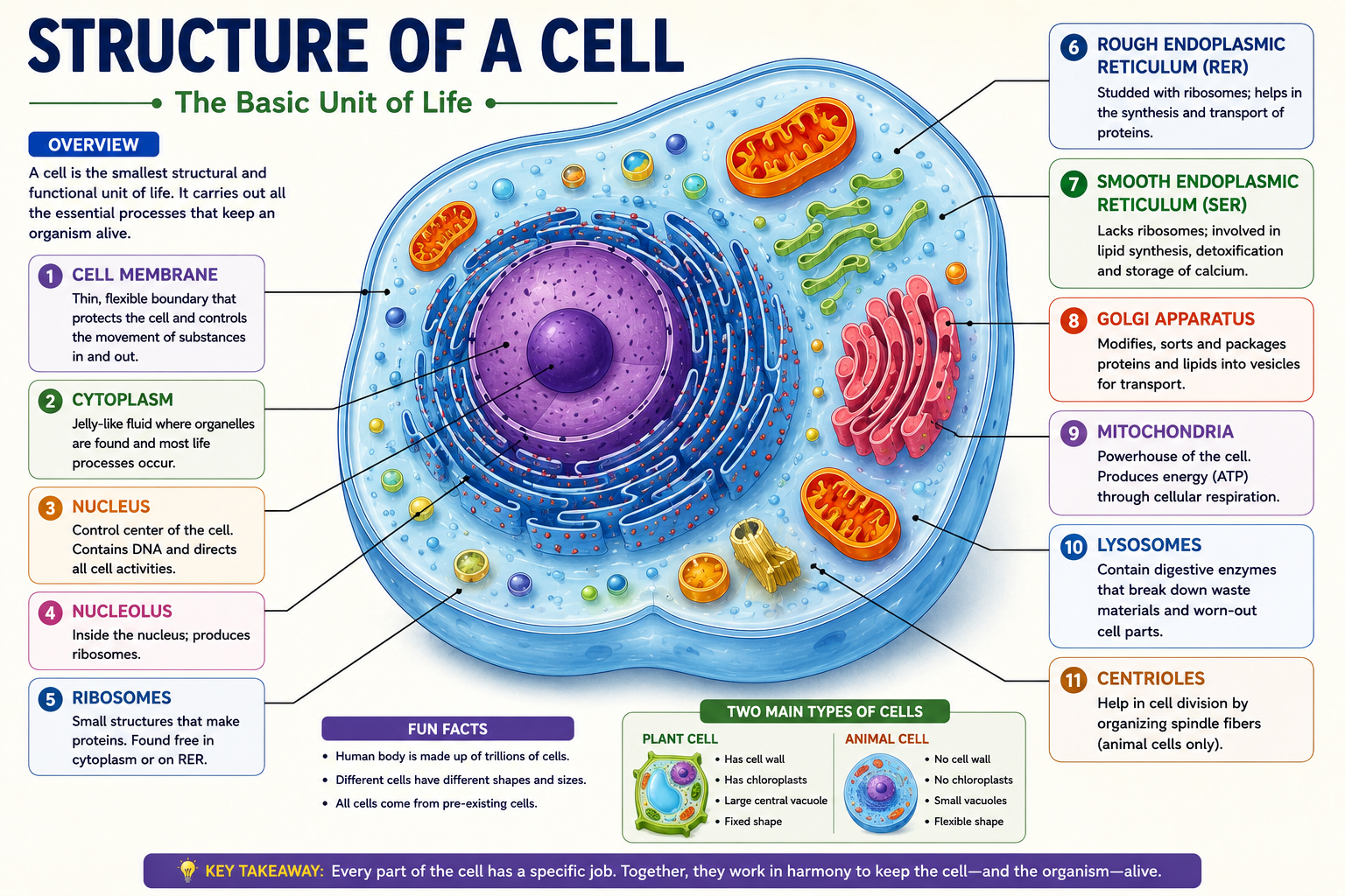
Now we get to the heart of it. Let’s walk through every major cell component, understanding not just what it looks like but why it’s built the way it is and what it actually does.
Cell Membrane
The cell membrane (plasma membrane) is the boundary that separates the cell’s interior from its external environment. Every cell has one. It’s a fluid mosaic—a dynamic, flexible structure composed of a phospholipid bilayer embedded with proteins, cholesterol, and carbohydrates.
The phospholipid bilayer is ingeniously designed. Each phospholipid molecule has a hydrophilic (water-loving) head and two hydrophobic (water-fearing) fatty acid tails. In an aqueous environment, these molecules spontaneously arrange themselves into a double layer, heads facing outward toward water, tails facing inward away from it. No construction required—thermodynamics does the work.
Functions of the cell membrane:
- Acts as a selective barrier, controlling what enters and exits the cell
- Contains receptor proteins that detect chemical signals
- Facilitates cell-to-cell recognition through surface carbohydrates
- Mediates cell adhesion and communication
Membrane proteins are embedded throughout and serve as channels, pumps, receptors, enzymes, and structural anchors. Their diversity is what makes the membrane’s selective permeability possible.
Cell Wall
The cell wall is a rigid outer layer found in plants, fungi, and prokaryotes—but NOT in animal cells. In plants, it’s composed primarily of cellulose microfibrils. In fungi, it’s made of chitin. In bacteria, it’s peptidoglycan.
The cell wall provides structural support, gives the cell its shape, prevents excessive water uptake (which could cause the cell to burst), and in plants, contributes to the upright growth habit through turgor pressure against the wall.
Cytoplasm
The cytoplasm is everything inside the cell membrane except the nucleus. It consists of:
- Cytosol – The liquid component; mostly water with dissolved salts, sugars, and proteins
- Organelles – Membrane-bound structures performing specific functions
- Cytoskeleton – A framework of protein filaments
The cytoplasm is far from an inert liquid. It’s the site of numerous metabolic reactions, including glycolysis (the first stage of cellular respiration), and it provides the medium through which materials move within the cell.
Nucleus
The nucleus is the cell’s command center—the largest organelle in most eukaryotic cells. It contains the cell’s DNA, organized into chromosomes, and controls all cellular activity by directing which proteins are made and when.
Structural components:
- Nuclear envelope – A double membrane with pores (nuclear pores) that regulate the movement of molecules between the nucleus and cytoplasm
- Nucleoplasm – The fluid inside the nucleus
- Chromatin – DNA associated with histone proteins; condenses into visible chromosomes during cell division
- Nucleolus – A region within the nucleus responsible for ribosome synthesis
Nucleolus
The nucleolus is not a membrane-bound organelle—it’s a dense, specialized region within the nucleus where ribosomal RNA (rRNA) is synthesized and ribosome subunits are assembled. It disappears during cell division when chromosomes condense, and reforms afterward. Cells that are highly active in protein synthesis (like liver cells and neurons) typically have large, prominent nucleoli.
Ribosomes
Ribosomes are the protein factories of the cell—the sites where messenger RNA (mRNA) is translated into protein. They’re not membrane-bound, which makes them unusual among organelles. They’re found in all cell types, both prokaryotic and eukaryotic, though their size differs (70S in prokaryotes, 80S in eukaryotes).
Ribosomes can be:
- Free ribosomes – Floating in the cytosol; synthesize proteins used within the cell
- Bound ribosomes – Attached to the rough endoplasmic reticulum; synthesize proteins destined for secretion or for the cell membrane
The size difference between prokaryotic and eukaryotic ribosomes is clinically significant—many antibiotics (streptomycin, erythromycin, tetracycline) work by binding to 70S prokaryotic ribosomes and disrupting protein synthesis without affecting the 80S ribosomes of human cells.
Endoplasmic Reticulum (Rough & Smooth)
The endoplasmic reticulum (ER) is an extensive network of interconnected membranes continuous with the outer nuclear membrane. Think of it as the cell’s internal highway system and manufacturing complex.
Rough ER:
- Studded with ribosomes on its outer surface (giving it the “rough” appearance)
- Site of synthesis of membrane proteins and secretory proteins
- Proteins are threaded into the ER lumen as they’re made, where they undergo folding and initial modifications
- Packages proteins into transport vesicles for the Golgi apparatus
Smooth ER:
- No ribosomes—smooth membrane surface
- Synthesizes lipids including phospholipids and steroid hormones
- In liver cells, contains enzymes that detoxify drugs, alcohol, and metabolic waste
- In muscle cells, stores calcium ions that trigger muscle contraction
- Plays a role in carbohydrate metabolism
Golgi Apparatus
The Golgi apparatus (or Golgi complex) is the cell’s post office and packaging center. It receives proteins from the rough ER via transport vesicles, modifies them further (adding sugar groups, cleaving signal sequences), sorts them, and packages them into vesicles for delivery to their final destinations.
Structurally, it consists of a stack of flattened membrane-bound sacs called cisternae. Materials enter from the cis face (receiving side, facing the ER) and exit from the trans face (shipping side, facing the cell membrane or lysosomes).
Destinations for Golgi-processed proteins:
- Secretion outside the cell (enzymes, hormones, antibodies)
- Lysosomes
- The cell membrane itself
Mitochondria
Mitochondria are the powerhouses of the cell—the sites of cellular respiration where glucose and other fuel molecules are converted into ATP (adenosine triphosphate), the cell’s universal energy currency.
Structure:
- Outer membrane – Smooth; permeable to most small molecules
- Inner membrane – Highly folded into projections called cristae, which dramatically increase surface area for ATP production
- Intermembrane space – Between the two membranes; site of proton accumulation during electron transport
- Matrix – The fluid-filled interior; contains enzymes for the Krebs cycle, mitochondrial DNA, and ribosomes
The presence of their own DNA and 70S ribosomes suggests mitochondria evolved from free-living bacteria that were engulfed by early eukaryotic cells—the endosymbiotic theory. This is one of the most well-supported hypotheses in evolutionary cell biology.
Cells with high energy demands (heart muscle cells, liver cells, neurons) have the most mitochondria—some heart muscle cells contain thousands.
Lysosomes
Lysosomes are the cell’s recycling centers and waste disposal units. They’re membrane-bound organelles containing approximately 50 different hydrolytic enzymes capable of breaking down proteins, nucleic acids, lipids, and carbohydrates.
The lysosomal membrane maintains an interior pH of around 5—significantly more acidic than the cytosol (pH ~7.2). This acidic environment is optimal for lysosomal enzymes and also serves as a safety mechanism: if a lysosome ruptures, its enzymes are largely inactive at the cytosol’s neutral pH.
Functions:
- Intracellular digestion of materials brought in by endocytosis
- Autophagy—breaking down damaged or worn-out organelles for recycling
- Programmed cell death (apoptosis) in some contexts
Clinically important: Lysosomal storage diseases (like Tay-Sachs disease) result from deficiencies in specific lysosomal enzymes, causing harmful accumulation of undigested substrates.
Peroxisomes
Peroxisomes are small, membrane-bound organelles containing enzymes that oxidize various organic molecules. A key byproduct of these reactions is hydrogen peroxide (H₂O₂)—potentially toxic—which is immediately broken down by catalase (also present in peroxisomes) into water and oxygen.
In liver cells, peroxisomes are particularly abundant and important for detoxifying alcohol and other harmful compounds. They’re also involved in fatty acid oxidation.
Vacuoles
Vacuoles are membrane-bound storage compartments. Their size and function vary dramatically between cell types:
- Plant cells have one large central vacuole that occupies up to 90% of cell volume, storing water (maintaining turgor pressure), ions, sugars, waste products, and pigments (the red and purple colors of many flowers and autumn leaves come from anthocyanins stored in vacuoles)
- Animal cells have small, temporary vacuoles used for food digestion (food vacuoles) or water regulation (contractile vacuoles in protists)
- Prokaryotes lack true vacuoles, though some have gas vesicles for buoyancy
Chloroplasts
Found only in plant cells and some protists, chloroplasts are the organelles where photosynthesis takes place. Like mitochondria, they have their own DNA and 70S ribosomes—strong evidence for endosymbiotic origin from photosynthetic bacteria (specifically cyanobacteria).
Structure:
- Outer membrane – Permeable to most small molecules
- Inner membrane – Less permeable; regulates material transport
- Thylakoids – Flattened membrane sacs organized into stacks called grana; site of light-dependent reactions of photosynthesis; contain chlorophyll
- Stroma – The fluid-filled space surrounding thylakoids; site of the Calvin cycle (light-independent reactions)
Cytoskeleton
The cytoskeleton is a dynamic network of protein filaments extending throughout the cytoplasm. It’s anything but static—it continuously assembles and disassembles, giving cells their shape, enabling movement, and organizing the interior.
Three major components:
- Microfilaments (actin filaments) – Thinnest; involved in cell movement, muscle contraction, and cell division
- Intermediate filaments – Medium thickness; provide mechanical strength; anchor organelles; include keratin in skin cells
- Microtubules – Thickest; form the spindle fibers during cell division; serve as tracks for motor proteins transporting organelles; form cilia and flagella
Centrioles
Centrioles are cylindrical structures made of microtubule triplets, found in animal cells (and some lower plants) but absent in most plant cells. Pairs of centrioles form the centrosome—the cell’s main microtubule-organizing center.
During cell division, centrioles help organize the mitotic spindle that pulls chromosomes to opposite poles of the dividing cell. They’re also the basal bodies from which cilia and flagella grow.
Cell Organelles and Their Functions (Complete Table)
| Organelle | Found In | Primary Function |
|---|---|---|
| Cell membrane | All cells | Selective barrier; cell signaling; transport regulation |
| Cell wall | Plants, fungi, bacteria | Structural support; shape; protection |
| Nucleus | Eukaryotes | Contains DNA; controls gene expression |
| Nucleolus | Eukaryotes | Synthesizes rRNA; assembles ribosome subunits |
| Ribosomes | All cells | Protein synthesis |
| Rough ER | Eukaryotes | Synthesis and processing of secretory proteins |
| Smooth ER | Eukaryotes | Lipid synthesis; detoxification; Ca²⁺ storage |
| Golgi apparatus | Eukaryotes | Modifies, sorts, and packages proteins |
| Mitochondria | Eukaryotes | ATP production via cellular respiration |
| Lysosomes | Animal cells mainly | Intracellular digestion; autophagy |
| Peroxisomes | Eukaryotes | Oxidative reactions; detoxification |
| Vacuoles | Eukaryotes | Storage; turgor pressure (plants) |
| Chloroplasts | Plants, some protists | Photosynthesis |
| Cytoskeleton | Eukaryotes | Cell shape; movement; organelle positioning |
| Centrioles | Animal cells (mostly) | Spindle formation during cell division |
Plant Cell vs Animal Cell
| Feature | Plant Cell | Animal Cell |
|---|---|---|
| Cell wall | Present (cellulose) | Absent |
| Chloroplasts | Present | Absent |
| Large central vacuole | Present | Absent (small vacuoles only) |
| Centrioles | Absent in most | Present |
| Shape | Regular, rectangular | Irregular, variable |
| Lysosomes | Rare | Common |
| Energy storage | Starch | Glycogen |
| Plasmodesmata | Present | Absent |
| Cilia/Flagella | Rare | Present in some |
| Endoplasmic reticulum | Present | Present |
Movement of Materials Across the Cell Membrane
Getting the right materials into a cell and waste products out is one of the cell’s most critical ongoing tasks. There are six major mechanisms—understanding each one, including whether it requires energy, is essential for any biology exam.
Diffusion
Diffusion is the net movement of molecules from an area of high concentration to an area of low concentration—down the concentration gradient. It requires no energy (passive process) and results from the random thermal motion of molecules.
Example: Oxygen diffuses from the high oxygen concentration in alveoli (air sacs in lungs) into red blood cells where oxygen concentration is lower. Carbon dioxide diffuses in the opposite direction.
Factors affecting diffusion rate:
- Concentration gradient (steeper = faster)
- Temperature (higher = faster)
- Molecular size (smaller = faster)
- Distance (shorter = faster)
- Surface area (larger = faster)
Osmosis
Osmosis is a special case of diffusion—specifically the net movement of water molecules across a selectively permeable membrane, from an area of lower solute concentration (higher water concentration) to an area of higher solute concentration (lower water concentration).
Key terms:
- Hypotonic solution – Lower solute concentration than the cell; water moves INTO the cell; animal cells may burst (lyse); plant cells become turgid
- Hypertonic solution – Higher solute concentration than the cell; water moves OUT of the cell; animal cells shrivel (crenate); plant cells become plasmolyzed
- Isotonic solution – Equal solute concentration; no net water movement
Example: Red blood cells placed in distilled water swell and eventually burst because water moves in by osmosis. The same cells in a concentrated salt solution shrivel because water moves out.
Facilitated Diffusion
Some molecules—glucose, amino acids, large ions—are too large or too charged to simply diffuse across the lipid bilayer. They move down their concentration gradient using transport proteins (channel proteins or carrier proteins) embedded in the membrane.
This is still a passive process—no energy required—but it requires specific protein facilitators. The rate is limited by the number of available transport proteins, which is why facilitated diffusion can become saturated (unlike simple diffusion).
Active Transport
Active transport moves molecules against their concentration gradient—from low concentration to high concentration. This requires energy (ATP) and specific carrier proteins (pumps).
Classic example: The sodium-potassium pump (Na⁺/K⁺ ATPase) found in virtually all animal cells. It actively pumps 3 sodium ions out of the cell and 2 potassium ions in, maintaining the electrochemical gradient essential for nerve impulse transmission and muscle contraction. For every cycle, one ATP molecule is consumed.
Active transport is essential for:
- Nerve and muscle function
- Nutrient absorption in the gut
- Kidney function (reabsorption of glucose and ions)
- Maintaining resting membrane potential in neurons
Endocytosis
Endocytosis is the process by which cells engulf large molecules or particles by wrapping the cell membrane around them, forming a vesicle that enters the cell. Three types:
- Phagocytosis (“cell eating”) – Cell engulfs large solid particles (bacteria, debris); used by immune cells (macrophages, neutrophils) to destroy pathogens
- Pinocytosis (“cell drinking”) – Cell engulfs extracellular fluid and dissolved solutes in small vesicles
- Receptor-mediated endocytosis – Highly specific; target molecules bind to receptor proteins in coated pits on the membrane surface, triggering vesicle formation; how cells take up cholesterol (via LDL receptors) and some hormones
Exocytosis
Exocytosis is the reverse of endocytosis—vesicles inside the cell fuse with the cell membrane and release their contents to the outside. This is how cells secrete products:
- Neurons release neurotransmitters by exocytosis
- Pancreatic cells secrete insulin and digestive enzymes
- Cells of the immune system release antibodies
- The Golgi apparatus uses exocytosis to deliver proteins to the cell membrane
Cell Communication
Cells don’t operate in isolation—they constantly send and receive chemical signals that coordinate their activities. This is called cell signaling or cell communication.
Three stages of cell signaling:
- Reception – A signaling molecule (ligand) binds to a receptor protein, usually on the cell surface
- Transduction – The binding event triggers a cascade of molecular changes inside the cell (signal transduction pathway)
- Response – The cell changes its behavior—activating genes, altering metabolism, triggering division, or initiating apoptosis
Types of signaling:
- Paracrine signaling – Signal affects nearby cells (growth factors in tissue repair)
- Endocrine signaling – Hormones travel through the bloodstream to distant target cells (insulin from the pancreas acting on muscle and liver cells)
- Synaptic signaling – Neurotransmitters cross synapses between neurons
- Autocrine signaling – A cell signals to itself
Cell signaling failures are at the heart of many diseases. Cancer often involves mutations in signaling proteins that cause cells to divide uncontrollably.
Cell Growth and Development
Individual cells grow between divisions, doubling most of their contents before dividing to produce two daughter cells. But in multicellular organisms, cell growth and development involves much more than simple enlargement.
Cell differentiation is the process by which genetically identical cells become structurally and functionally distinct. A skin cell, a neuron, and a liver cell all contain identical DNA, but they express different genes and consequently have different structures and perform different functions.
Differentiation is controlled by:
- Gene expression patterns – Which genes are activated or silenced
- Transcription factors – Proteins that bind to DNA and control transcription
- Epigenetic modifications – Chemical changes to DNA or histones that affect gene expression without changing the DNA sequence
- Signaling from neighboring cells – Chemical signals from adjacent cells influence which genes are expressed
Cell Cycle Overview
The cell cycle is the ordered sequence of events through which a cell grows and divides. It’s divided into two major phases: interphase and the mitotic phase.
Interphase
Interphase is the longest phase of the cell cycle—the period between cell divisions when the cell is actively growing, carrying out its normal functions, and preparing for division. It consists of three subphases:
- G₁ (Gap 1) – Cell grows in size; produces proteins and organelles; a checkpoint (G₁ checkpoint) determines whether conditions are favorable for division
- S phase (Synthesis) – DNA replication occurs; each chromosome is duplicated to form two identical sister chromatids joined at the centromere
- G₂ (Gap 2) – Cell continues to grow; produces proteins needed for mitosis; another checkpoint (G₂ checkpoint) verifies DNA replication accuracy
Some cells exit the cell cycle and enter a non-dividing state called G₀. Most neurons in the adult brain are in G₀ permanently.
Mitosis
Mitosis is the division of the nucleus, producing two genetically identical daughter nuclei. It has four distinct phases:
- Prophase – Chromatin condenses into visible chromosomes; spindle fibers begin to form; nuclear envelope breaks down
- Metaphase – Chromosomes align along the cell’s equatorial plate (metaphase plate); spindle fibers attach to centromeres. This is the easiest phase to count chromosomes.
- Anaphase – Sister chromatids are pulled apart to opposite poles of the cell; cell elongates
- Telophase – Chromosomes reach the poles and begin to decondense; nuclear envelopes reform around each set of chromosomes; spindle fibers disappear
Memory aid: PMAT — Prophase, Metaphase, Anaphase, Telophase
Cytokinesis
Cytokinesis is the division of the cytoplasm, producing two separate daughter cells. It typically begins during late anaphase and completes after telophase.
In animal cells: a contractile ring of actin filaments pinches the cell in two (cleavage furrow)
In plant cells: membrane vesicles from the Golgi fuse along the cell’s midline to form a cell plate, which develops into a new cell wall between the daughter cells
Cell Division and Its Importance
Cell division is essential for three fundamental biological processes:
- Growth – Multicellular organisms grow by producing more cells through mitosis
- Repair and replacement – Damaged or dead cells are replaced by new ones; your skin cells are completely replaced approximately every 2–4 weeks
- Reproduction – Asexual reproduction in single-celled organisms occurs through cell division; sexual reproduction requires meiosis to produce gametes
Meiosis (distinct from mitosis) produces four genetically diverse haploid daughter cells—sperm and egg cells. It involves two rounds of division (Meiosis I and II) and includes crossing over, which shuffles genetic material between homologous chromosomes, generating genetic variation.
When cell division is uncontrolled—when the checkpoints of the cell cycle fail—cancer results. Tumor suppressor genes (like p53) normally enforce checkpoints and trigger apoptosis in damaged cells. When these genes are mutated, the brakes on cell division are lost.
How Cells Produce Energy
Cellular Respiration
Cellular respiration is the process by which cells break down glucose to produce ATP. It occurs in three stages:
- Glycolysis (in the cytoplasm)
- Glucose (6 carbon) is split into two pyruvate molecules (3 carbon each)
- Produces 2 ATP and 2 NADH net per glucose
- Doesn’t require oxygen—can occur anaerobically
- Krebs Cycle / Citric Acid Cycle (in the mitochondrial matrix)
- Pyruvate is converted to acetyl-CoA, which enters the cycle
- Each turn produces CO₂, NADH, FADH₂, and 1 ATP
- The cycle turns twice per glucose molecule
- Electron Transport Chain / Oxidative Phosphorylation (inner mitochondrial membrane)
- NADH and FADH₂ donate electrons to the chain
- Electrons move through protein complexes, releasing energy
- Energy is used to pump H⁺ ions across the membrane, creating a gradient
- H⁺ flows back through ATP synthase, producing ATP (approximately 28–34 ATP per glucose)
- Oxygen is the final electron acceptor, forming water
Overall equation:
C₆H₁₂O₆ + 6O₂ → 6CO₂ + 6H₂O + ~36-38 ATPATP Production
ATP (adenosine triphosphate) is the cell’s primary energy currency. When ATP is hydrolyzed to ADP + phosphate, energy is released and used to drive cellular work—muscle contraction, active transport, biosynthesis, and more.
The body continuously recycles ATP—at rest, a person turns over approximately 40 kg of ATP per day. During intense exercise, that rate increases dramatically.
Photosynthesis in Plant Cells
While animal cells can only extract energy from organic molecules, plant cells can capture energy directly from sunlight through photosynthesis—one of the most important reactions on Earth.
Overall equation:
6CO₂ + 6H₂O + Light Energy → C₆H₁₂O₆ + 6O₂Photosynthesis occurs in chloroplasts in two stages:
- Light-dependent reactions (thylakoid membranes): Light energy is captured by chlorophyll; water is split releasing O₂; ATP and NADPH are produced
- Calvin cycle (stroma): ATP and NADPH drive the fixation of CO₂ into glucose
The relationship between photosynthesis and cellular respiration is complementary—photosynthesis captures energy and stores it in glucose; cellular respiration releases that energy as ATP. The oxygen produced by photosynthesis is used in cellular respiration; the CO₂ produced by respiration is used in photosynthesis. Life on Earth runs on this elegant cycle.
Cell Specialization
In multicellular organisms, cells become specialized—structurally and functionally tailored for specific tasks. This specialization allows organisms to achieve levels of complexity and efficiency impossible for single cells.
Examples of specialized cells and their adaptations:
| Cell Type | Organism | Structural Adaptation | Function |
|---|---|---|---|
| Red blood cell | Humans | Biconcave shape; no nucleus; packed with hemoglobin | Oxygen transport |
| Neuron | Humans | Long axon; branching dendrites | Signal transmission |
| Guard cell | Plants | Contains chloroplasts; changes shape with water content | Controls stomatal opening |
| Sperm cell | Animals | Flagellum; many mitochondria; streamlined shape | Fertilization |
| Root hair cell | Plants | Long projection; no chloroplasts | Water and mineral absorption |
| Muscle cell (myocyte) | Animals | Long, striated fibers; abundant mitochondria | Contraction |
Cell specialization means that not all cells do all jobs—but every cell type is essential. This is why organ failure is serious: losing a population of specialized cells can be catastrophic for the whole organism.
Cell Structure in Different Organisms
Cell biology isn’t one-size-fits-all. Structure varies meaningfully across major groups of life:
Bacterial cells: Simple prokaryotic cells with no membrane-bound organelles. Some have flagella for movement. Cell walls of peptidoglycan are the target of penicillin-class antibiotics.
Archaeal cells: Prokaryotic like bacteria but biochemically distinct. Many are extremophiles. Their cell membranes contain unique lipids that provide stability in extreme conditions.
Fungal cells: Eukaryotic with chitin cell walls. Yeasts are unicellular; molds and mushrooms are multicellular. No chloroplasts.
Plant cells: Eukaryotic with cellulose cell walls, chloroplasts, and large central vacuoles. Two types: parenchyma (metabolically active), collenchyma (support), sclerenchyma (rigid support).
Animal cells: Eukaryotic without cell walls. Highly diverse specialization. Centrioles prominent.
Protist cells: Enormously diverse eukaryotic cells. Some photosynthesize (algae); some are heterotrophic (amoeba); some are parasitic (Plasmodium, causing malaria).
Common Cell Biology Terms Every Student Should Know
| Term | Definition |
|---|---|
| Organelle | A specialized structure within a cell that performs a specific function |
| Selectively permeable | Allows only certain substances to pass through |
| Concentration gradient | The difference in concentration of a substance between two areas |
| Turgor pressure | Pressure exerted by water in the vacuole against the cell wall |
| Apoptosis | Programmed cell death |
| Endosymbiosis | Theory that mitochondria and chloroplasts originated as free-living bacteria |
| Cell plate | Structure formed during cytokinesis in plant cells |
| Checkpoint | Control point in the cell cycle where conditions are assessed before proceeding |
| Differentiation | Process by which a cell becomes specialized |
| Cytosol | The liquid component of cytoplasm |
| Fluid mosaic model | Model describing the cell membrane as a dynamic bilayer with embedded proteins |
| Electrochemical gradient | Combination of concentration gradient and electrical charge difference |
| Lysis | Rupture of a cell due to excess water uptake |
| Plasmolysis | Shrinkage of cytoplasm from cell wall due to water loss in a hypertonic environment |
| Semi-conservative replication | DNA replication method where each new double helix contains one old and one new strand |
Common Mistakes Students Make
These are the errors I see most consistently from students, year after year. Knowing them before your exam is a genuine advantage.
1. Confusing osmosis with diffusion
Diffusion refers to the movement of any molecule from high to low concentration. Osmosis is specifically the movement of water across a selectively permeable membrane. Water moves from where there’s less solute (more water) to where there’s more solute (less water). Students often get the direction backwards.
2. Saying the nucleus “controls” protein synthesis
The nucleus controls gene expression—it determines which proteins are made. But actual protein synthesis happens at ribosomes in the cytoplasm (or on the rough ER). The nucleus is the director; ribosomes are the factory floor.
3. Forgetting that plant cells have mitochondria
Students sometimes assume plant cells only photosynthesize and don’t respire. Plants respire continuously (24 hours a day). Photosynthesis only happens in light, but respiration never stops.
4. Claiming active transport moves molecules down a concentration gradient
Active transport always works against the concentration gradient—from low to high. If it’s going down the gradient, it’s passive (diffusion or facilitated diffusion). Active transport is the only mechanism that requires ATP.
5. Mixing up mitosis and meiosis
Mitosis produces 2 genetically identical diploid cells (for growth and repair). Meiosis produces 4 genetically diverse haploid cells (gametes). These are frequently confused on exams, especially regarding chromosome number.
6. Describing the Golgi as just “packaging”
The Golgi apparatus doesn’t just package—it modifies proteins (adding sugars, cleaving sequences) and sorts them to different destinations. This distinction matters on higher-level exams.
Best Tips to Study Cell Biology
1. Build an organelle master table yourself
Don’t just read the table in this guide—construct your own from memory, adding to it as you learn. The act of constructing it reinforces learning far more effectively than passive reading.
2. Use spatial thinking for membrane transport
Draw a cell membrane with “inside” and “outside” labeled. Then for each transport mechanism, draw arrows showing direction of movement and whether energy is required. Visual spatial understanding of transport prevents the confusion that trips up so many students.
3. Learn the endosymbiotic theory as a narrative
The idea that mitochondria and chloroplasts evolved from bacteria that were engulfed by early cells explains so many features at once: their double membranes, their own DNA, their 70S ribosomes, their similar size to bacteria. Learning it as a story makes the features memorable.
4. Practice drawing cell diagrams from scratch
Close your notes and draw a plant cell and an animal cell from memory, labeling every organelle. Do this repeatedly. Biology exams frequently include labeling questions, and drawing from memory is the fastest way to identify what you’ve actually retained versus what you’re just recognizing when you see it.
5. Connect everything to disease
Cancer = cell cycle dysfunction. Tay-Sachs = lysosomal enzyme deficiency. Familial hypercholesterolemia = LDL receptor defect affecting receptor-mediated endocytosis. Cystic fibrosis = defective chloride channel (transport protein) in cell membranes. Every cell biology concept connects to real disease, and those connections make the content stick.
6. Use the PMAT mnemonic for mitosis, but understand each phase
Knowing the acronym alone isn’t enough. Know what physically happens to chromosomes, spindle fibers, and the nuclear envelope at each stage. Exam questions often describe a phase without naming it and ask you to identify it.
Cell Structure and Function Practice Questions
20 Multiple Choice Questions with Answers
- Which organelle is responsible for producing ATP through cellular respiration?
- A) Chloroplast
- B) Ribosome
- C) Mitochondria ✓
- D) Golgi apparatus
- A cell placed in a hypotonic solution will:
- A) Shrink due to water leaving
- B) Remain unchanged
- C) Swell due to water entering ✓
- D) Undergo immediate apoptosis
- Which of the following is found in plant cells but NOT animal cells?
- A) Mitochondria
- B) Ribosomes
- C) Endoplasmic reticulum
- D) Chloroplasts ✓
- The fluid mosaic model describes:
- A) Cytoplasmic organization
- B) The structure of the cell membrane ✓
- C) DNA replication
- D) The Golgi apparatus
- Which phase of mitosis do chromosomes align at the cell’s equatorial plate?
- A) Prophase
- B) Anaphase
- C) Metaphase ✓
- D) Telophase
- Active transport differs from passive transport in that active transport:
- A) Requires a concentration gradient
- B) Uses protein channels
- C) Requires ATP energy ✓
- D) Only moves water molecules
- The nucleolus is the site of:
- A) DNA replication
- B) Protein synthesis
- C) rRNA synthesis and ribosome assembly ✓
- D) Lipid production
- Ribosomes in prokaryotic cells are:
- A) 80S type
- B) 70S type ✓
- C) Absent entirely
- D) Membrane-bound
- Which organelle modifies, sorts, and packages proteins for secretion?
- A) Rough ER
- B) Lysosome
- C) Golgi apparatus ✓
- D) Peroxisome
- DNA replication occurs during which phase of the cell cycle?
- A) G₁ phase
- B) S phase ✓
- C) G₂ phase
- D) M phase
- Cell plate formation during cytokinesis occurs in:
- A) Animal cells
- B) Bacterial cells
- C) Plant cells ✓
- D) Fungal cells
- Which membrane transport mechanism requires a carrier protein but no energy?
- A) Active transport
- B) Osmosis
- C) Phagocytosis
- D) Facilitated diffusion ✓
- The endosymbiotic theory proposes that mitochondria evolved from:
- A) Invaginations of the cell membrane
- B) Free-living bacteria ✓
- C) RNA molecules
- D) Condensed chromatin
- Lysosomes contain:
- A) Photosynthetic pigments
- B) ATP synthase
- C) Hydrolytic enzymes ✓
- D) Genetic material
- During which mitotic phase does the nuclear envelope reform?
- A) Prophase
- B) Metaphase
- C) Anaphase
- D) Telophase ✓
- The smooth endoplasmic reticulum is primarily responsible for:
- A) Protein synthesis with ribosomes
- B) Lipid synthesis and detoxification ✓
- C) ATP production
- D) rRNA synthesis
- Which type of endocytosis is used by white blood cells to engulf bacteria?
- A) Pinocytosis
- B) Receptor-mediated endocytosis
- C) Phagocytosis ✓
- D) Exocytosis
- The sodium-potassium pump is an example of:
- A) Passive diffusion
- B) Osmosis
- C) Active transport ✓
- D) Facilitated diffusion
- Which organelle contains its own DNA and ribosomes?
- A) Golgi apparatus
- B) Lysosome
- C) Vacuole
- D) Mitochondria ✓
- Cell differentiation refers to:
- A) The process of cell division
- B) Cell death by apoptosis
- C) Cells becoming structurally and functionally specialized ✓
- D) Movement of cells during development
10 Short Answer Questions
- Describe the fluid mosaic model of the cell membrane. What molecules make up this structure and what is the functional significance of its organization?
- Explain the difference between osmosis and diffusion. Give one biological example of each.
- What are the three principles of cell theory? Identify the scientist most closely associated with each principle.
- Describe three structural differences between prokaryotic and eukaryotic cells. Explain the functional consequence of each difference.
- Explain the role of the Golgi apparatus in protein processing and secretion. What is the difference between the cis and trans faces?
- Describe the stages of the cell cycle in order. What occurs at each G checkpoint and why are checkpoints important?
- Compare and contrast mitosis and meiosis. Include chromosome number, number of daughter cells, genetic diversity, and biological purpose.
- Explain the cohesion-tension theory—wait, that’s plant biology. Instead: explain the sodium-potassium pump. What moves in which direction, what energy source is used, and why is this pump physiologically critical?
- Describe the process of endocytosis and exocytosis. Give one specific biological example of each.
- What is cell specialization? Give three examples of specialized cells, describing the structural adaptation and its functional advantage.
5 Long Answer Questions
- Describe the structure and function of the cell membrane in detail. Explain the fluid mosaic model, describing the role of phospholipids, proteins, cholesterol, and carbohydrates. Then describe all six mechanisms by which materials cross the cell membrane, specifying which require energy, which require protein carriers, and the direction of movement relative to the concentration gradient. Use a specific biological example for at least four mechanisms.
- Compare prokaryotic and eukaryotic cells comprehensively. Address their size, genetic material organization, membrane-bound organelles, cell wall composition, ribosome type, and reproduction. Explain why the distinction between these cell types is fundamental to medicine, specifically in terms of how antibiotics selectively target bacteria without harming human cells.
- Describe the cell cycle in detail, including all phases of interphase and all phases of mitosis. Explain what occurs at each cell cycle checkpoint, what happens when checkpoint controls fail, and how this relates to cancer. Include a description of cytokinesis and how it differs between plant and animal cells.
- Explain how cells produce energy through cellular respiration. Describe the three stages (glycolysis, Krebs cycle, and electron transport chain), specifying where each occurs in the cell, what inputs are required, what outputs are produced, and approximately how much ATP is generated at each stage. Explain how this process relates to photosynthesis in plant cells.
- Discuss the endomembrane system of eukaryotic cells, including the nuclear envelope, endoplasmic reticulum (rough and smooth), Golgi apparatus, vesicles, lysosomes, and cell membrane. Trace the journey of a secretory protein from its synthesis to its release from the cell, explaining what happens to the protein at each location. Discuss how defects in this system can lead to human disease, using at least one specific example.
Cell Biology Revision Checklist
Go through this honestly before your exam. If you can’t check something off, go back and review that section.
- I can state and explain the three principles of cell theory and name the scientists behind each
- I can distinguish prokaryotic from eukaryotic cells across at least six features
- I can draw and label a plant cell and an animal cell with all major organelles
- I can describe the function of every organelle in the complete organelle table
- I can list five differences between plant and animal cells with functional explanations
- I can explain all six mechanisms of membrane transport and distinguish passive from active
- I can explain osmosis and correctly predict what happens to a cell in hypotonic, hypertonic, and isotonic solutions
- I can describe all four phases of mitosis and explain what physically happens in each
- I can distinguish mitosis from meiosis in terms of chromosome number, cell number, and purpose
- I can explain the three stages of cellular respiration and where each occurs in the cell
- I can describe cell cycle checkpoints and explain why they matter for cancer biology
- I can give three examples of specialized cells and describe their structural adaptations
- I can explain the endosymbiotic theory and the evidence supporting it
- I have completed at least 20 MCQs and 3 long answer questions from this guide
Best Books for Learning Cell Biology
- “Molecular Biology of the Cell” by Alberts, Johnson, Lewis, et al. – The definitive cell biology reference used in universities worldwide. If you want to understand cell biology at the deepest level, this is the book. The sixth edition is available online free through NCBI Bookshelf.
- “Cell Biology: A Short Course” by Bolsover, Shephard, White, and Bhatt – Shorter and more accessible than Alberts, making it ideal for students who need a solid but manageable introduction without getting lost in encyclopedic detail.
- Campbell Biology (any recent edition) – The gold-standard general biology textbook has exceptional chapters on cell structure, membrane transport, and cell division. Outstanding for AP Biology and equivalent examinations.
- “The Cell: A Very Short Introduction” by Terence Allen and Graham Cowling – An excellent, compact read for students who want conceptual clarity before diving into detailed textbooks. Reads almost like a story of how cell biology developed.
- “Lehninger Principles of Biochemistry” by Nelson and Cox – For students who want to go deeper on the biochemical side of cell function—ATP production, enzyme kinetics, membrane biochemistry. More demanding but richly rewarding.
Free Online Cell Biology Resources
- OpenStax Biology 2e – Cell Chapter – Free, peer-reviewed, comprehensive. Covers cell structure, membrane transport, and cell reproduction with clear diagrams and review questions.
- Khan Academy – Cell Biology – Excellent video lessons covering every major cell biology topic with integrated practice questions. Perfect for visual learners and AP Biology preparation.
- Biology LibreTexts – Cell Biology – Open-access academic content organized clearly by topic. Suitable for both introductory and advanced study at university level.
- HHMI BioInteractive – Cell Biology Resources – Research-quality educational animations and videos from the Howard Hughes Medical Institute. The 3D animations of cellular processes are genuinely outstanding.
- NCBI Bookshelf – Molecular Biology of the Cell – The full text of Alberts’ comprehensive cell biology textbook, freely accessible online. One of the most authoritative resources in the field.
Frequently Asked Questions
1. What is the basic unit of life?
The cell is the basic structural and functional unit of all living organisms. Every living thing is composed of one or more cells, and all life processes occur within cells or are carried out by cells.
2. What is the difference between a prokaryotic and a eukaryotic cell?
Prokaryotic cells lack a membrane-bound nucleus and membrane-bound organelles. Eukaryotic cells have a true nucleus enclosed by a membrane and contain specialized organelles. Bacteria and archaea are prokaryotes; plants, animals, fungi, and protists are eukaryotes.
3. What is the most important organelle in a cell?
This depends on what you mean by “important”—every organelle is essential for cell survival. The nucleus is often considered most critical because it contains the genetic information controlling all cell activities. The mitochondria, however, are indispensable for energy production.
4. Why do animal cells not have cell walls?
Animal cells don’t need rigid cell walls because they’re supported by internal cytoskeletal structures and the tissues of which they’re part. Cell walls also limit movement and flexibility—qualities many animal cells (like white blood cells and sperm) require.
5. What is the difference between diffusion and osmosis?
Diffusion is the net movement of any substance from high to low concentration. Osmosis is specifically the diffusion of water across a selectively permeable membrane—from lower solute concentration to higher solute concentration.
6. What happens to a cell in a hypertonic solution?
Water moves out of the cell by osmosis (toward the higher solute concentration outside). Animal cells shrivel and crenate. Plant cells lose turgor and undergo plasmolysis as the cytoplasm shrinks away from the cell wall.
7. How is meiosis different from mitosis?
Mitosis produces two genetically identical diploid daughter cells for growth and repair. Meiosis produces four genetically unique haploid daughter cells (gametes) for sexual reproduction. Meiosis includes two rounds of division and crossing over between homologous chromosomes.
8. What is the role of ATP in the cell?
ATP (adenosine triphosphate) is the cell’s primary energy currency. Energy stored in ATP is released when it’s hydrolyzed to ADP + phosphate. This released energy powers active transport, muscle contraction, biosynthesis, and virtually every energy-requiring cellular process.
9. Why is the cell membrane described as “selectively permeable”?
The cell membrane allows some substances to pass freely (small nonpolar molecules like oxygen and CO₂) while blocking others (large molecules, charged ions) unless they use specific transport proteins. This selectivity allows cells to maintain a controlled internal environment.
10. What is apoptosis and why is it important?
Apoptosis is programmed cell death—a controlled, orderly process by which damaged, infected, or unnecessary cells are dismantled and their components recycled. It’s essential for development (sculpting fingers by eliminating cells between them), immune function, and cancer prevention.
11. What is the endosymbiotic theory?
The endosymbiotic theory proposes that mitochondria and chloroplasts evolved from free-living bacteria that were engulfed by early eukaryotic cells. Evidence includes their double membranes, own circular DNA, 70S ribosomes, and reproduction by binary fission—all characteristics of bacteria.
12. How does the Golgi apparatus know where to send proteins?
Proteins are tagged with specific signal sequences—short amino acid chains that act as molecular addresses. The Golgi reads these tags and sorts proteins into vesicles destined for secretion, the cell membrane, or lysosomes. This sorting mechanism is highly specific and essential for cell organization.
Summary
This complete guide to cell structure and function has taken you from the first observations of cells through the sophisticated molecular machinery that keeps every cell alive. Here are the essential threads to hold onto.
Cells are the fundamental units of life—all living things are composed of cells, and all cells come from pre-existing cells (cell theory). They fall into two broad categories: prokaryotic (no nucleus, no membrane-bound organelles—bacteria and archaea) and eukaryotic (true nucleus, membrane-bound organelles—plants, animals, fungi, protists).
Each organelle has a specific, irreplaceable function. The nucleus stores and expresses genetic information. Mitochondria produce ATP. Chloroplasts conduct photosynthesis. The rough ER and Golgi apparatus work together to produce and deliver proteins. Lysosomes recycle cellular waste. The cytoskeleton provides structure and enables movement.
Materials cross the cell membrane through six mechanisms ranging from passive diffusion to energy-requiring active transport and vesicle-mediated endocytosis and exocytosis. The cell cycle—interphase followed by mitosis and cytokinesis—precisely controls cell division, with checkpoints preventing errors that could lead to cancer.
In multicellular organisms, cells specialize for specific functions, each structurally adapted to perform its role with maximum efficiency. Understanding cell structure and function is understanding the foundation upon which all biological complexity is built.
Final Thoughts
The cell is where biology becomes real. Everything that happens in living organisms—growth, disease, healing, reproduction, inheritance—happens in and between cells. Once you truly understand how a cell is structured and what each component does, the rest of biology starts to make logical sense in a way it simply can’t without that foundation.
Students who invest time in genuinely understanding cell biology—not just memorizing organelle names but understanding why each structure is built the way it is—find that their understanding of genetics, physiology, microbiology, and medicine all deepen simultaneously. It’s the best return on investment in all of biology education.
Use the revision checklist. Work through the practice questions without looking at the answers until you’ve genuinely tried. Draw your cell diagrams until they feel natural. And connect everything—every organelle, every transport mechanism, every phase of mitosis—to something in the real world.
The more clearly you understand cells, the more clearly you’ll understand life itself.
Good luck with your studies.
References
- OpenStax Biology 2e – Cell Chapters – openstax.org/books/biology-2e
- Khan Academy – AP Biology: Cell Structure and Function – khanacademy.org
- Biology LibreTexts – Cell Biology – bio.libretexts.org
- HHMI BioInteractive – Cell Biology – biointeractive.org
- NCBI Bookshelf – Molecular Biology of the Cell – ncbi.nlm.nih.gov/books
Article Disclaimer
This article is intended for educational and informational purposes only. While LearnMinto strives to provide accurate, well-researched, and up-to-date information, cell biology is a dynamic field of science that continues to evolve through ongoing research and discoveries. Readers should verify important academic concepts through official textbooks, educational institutions, examination boards, or trusted scientific resources before relying on this content for exams or academic purposes. LearnMinto is not affiliated with any specific school, university, research institution, or examination board. This content is designed solely to support learning, revision, and educational development and should not be considered medical or professional scientific advice.