
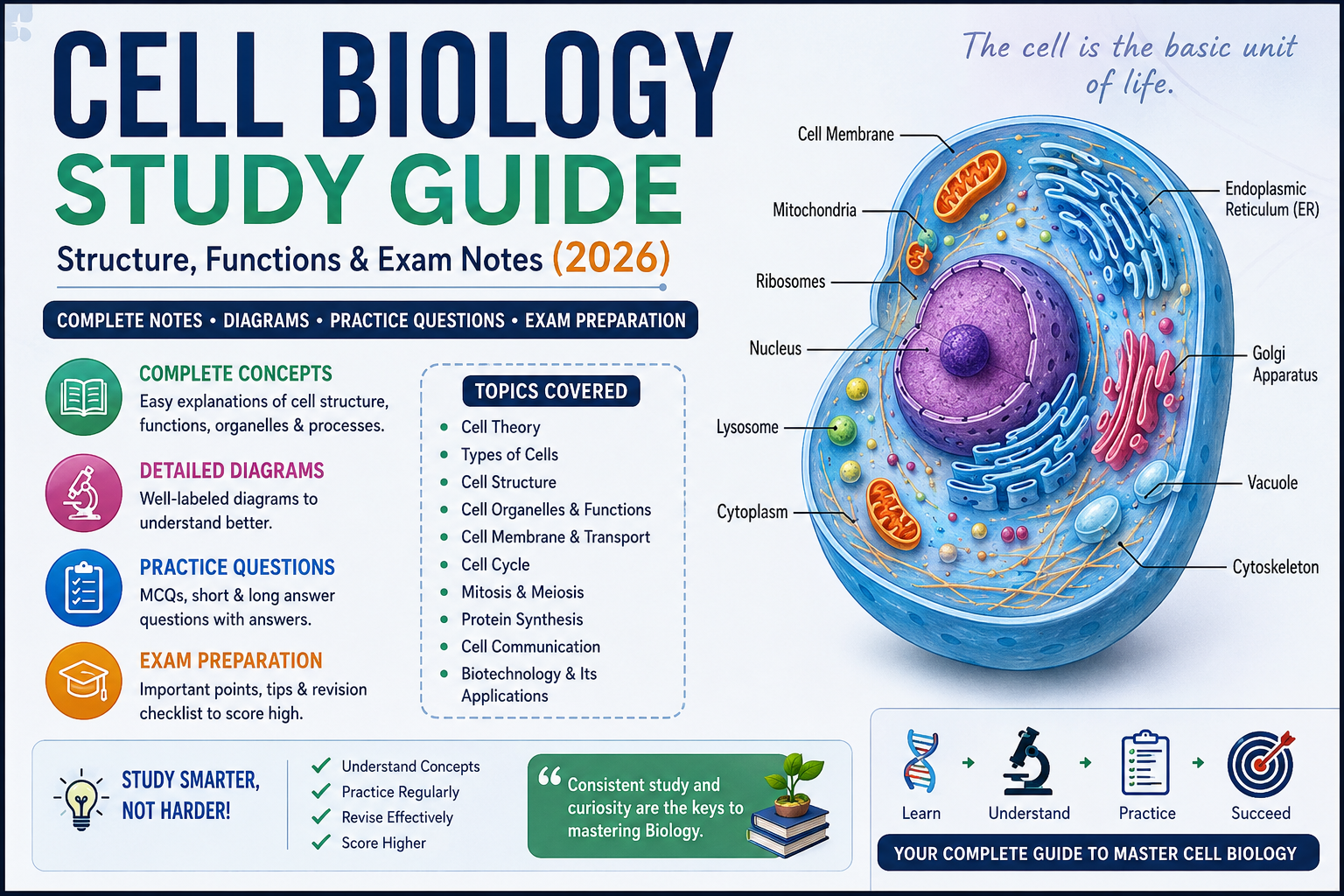
Here’s something worth thinking about: every living thing you’ve ever seen—every tree, every insect, every person—is built from cells. Cells so small that millions of them could fit on the head of a pin. Yet inside each one, there’s an entire world of organized activity happening every second of every day.
Cell biology is where the real story of life begins. It’s not just a topic you study to pass an exam. It’s the foundation of every other subject in biology. When you understand how cells work, genetics makes more sense, physiology becomes clearer, and even topics like evolution and ecology start to connect in ways they didn’t before.
This cell biology study guide is designed to be everything you need in one place. Whether you’re a high school student tackling this topic for the first time, a college student reviewing for finals, a teacher looking for organized notes, or someone preparing for AP Biology, IB Biology, GCSE, or MCAT exams, this guide walks you through every essential concept—clearly, thoroughly, and without unnecessary fluff.
Let’s start from the beginning and build up from there.
Key Takeaways
- All living organisms are made of one or more cells—this is the foundation of cell theory.
- There are two main types of cells: prokaryotic (no nucleus) and eukaryotic (with nucleus).
- Each organelle has a specific function, and understanding those functions is critical for exams.
- Materials cross cell membranes through diffusion, osmosis, facilitated diffusion, and active transport.
- Cell division occurs through mitosis (growth and repair) and meiosis (reproduction).
- Protein synthesis links DNA to function through transcription and translation.
- Drawing and labeling cell diagrams from memory is one of the highest-yield study strategies.
- This guide includes 35 practice questions across three levels for complete exam preparation.
What Is Cell Biology?
Cell biology—sometimes called cytology—is the branch of biology that studies the structure, function, behavior, and life cycle of cells. It focuses on understanding the cell as the basic unit of life and examines how cells carry out the processes that keep organisms alive.
This field explores questions like:
- What is a cell made of?
- How do cells produce energy?
- How do they reproduce?
- How do they communicate with each other?
- How do they respond to damage or disease?
Cell biology sits at the intersection of biochemistry, genetics, and physiology. It’s not isolated—it connects to virtually every other area of biological science. When researchers study cancer, they’re studying abnormal cell behavior. When pharmaceutical companies develop drugs, they’re targeting specific cellular processes. When genetic engineers use CRISPR, they’re operating at the cellular and molecular level.
For students, cell biology is typically one of the first major units in a biology course, and for good reason. Without understanding cells, nothing else in biology fully makes sense.
Why Is Cell Biology Important?
Understanding cell biology matters well beyond the classroom, and it’s worth taking a moment to appreciate why.
Medically, nearly every disease involves cellular malfunction. Cancer is uncontrolled cell division. HIV attacks specific immune cells. Diabetes involves problems with how cells respond to insulin. Alzheimer’s disease involves the death of neurons. Understanding cells is literally understanding disease.
Pharmaceutically, drug development depends on knowing how cells work. Antibiotics interfere with bacterial cell processes without harming human cells. Chemotherapy drugs target rapidly dividing cancer cells. None of this is possible without deep knowledge of cell biology.
Biotechnologically, innovations like gene therapy, stem cell research, and organ regeneration are all built on our understanding of how cells grow, differentiate, and function.
Ecologically, cells connect us to every other living organism. The photosynthetic cells in plants produce the oxygen we breathe. Microbial cells in soil recycle nutrients. Everything in an ecosystem runs on cellular processes.
For students specifically, cell biology forms the backbone of AP Biology, IB Biology, MCAT preparation, nursing school entrance exams, and virtually every biology-related degree program. Mastering it early gives you a significant advantage throughout your entire academic journey.
History of Cell Biology
Cell biology didn’t emerge fully formed. It developed gradually as technology and scientific thinking evolved over several centuries.
| Year | Scientist | Contribution |
|---|---|---|
| 1665 | Robert Hooke | First observed cells using a primitive microscope; named them “cells” after examining cork |
| 1674 | Antonie van Leeuwenhoek | First observed living cells (bacteria and protozoa) using improved lenses |
| 1831 | Robert Brown | Discovered the nucleus within plant cells |
| 1838 | Matthias Schleiden | Proposed that all plants are made of cells |
| 1839 | Theodor Schwann | Extended cell theory to animals |
| 1855 | Rudolf Virchow | Stated that all cells come from pre-existing cells (Omnis cellula e cellula) |
| 1879 | Walther Flemming | Discovered and described mitosis |
| 1953 | Watson & Crick | Described the double-helix structure of DNA |
| 1960s | Various researchers | Development of electron microscopy revealed ultrastructure of organelles |
| 1990s–present | Multiple researchers | Advances in molecular cell biology, genomics, and CRISPR technology |
This history matters because it shows that our understanding of cells evolved through observation, technology, and collaboration. It also reminds us that science builds on itself—each discovery opened doors to the next.
Cell Theory Explained
Cell theory is one of the most fundamental concepts in all of biology. It’s not just a definition to memorize—it’s a framework that explains how life is organized.
The three principles of cell theory are:
- All living organisms are composed of one or more cells.
Every life form, from bacteria to blue whales, is made entirely of cells. Viruses are the notable exception—they’re not made of cells, which is one reason scientists debate whether viruses are truly “alive.” - The cell is the basic unit of life.
The cell is the smallest unit capable of carrying out all the functions of life: obtaining energy, responding to stimuli, reproducing, and maintaining homeostasis. - All cells arise from pre-existing cells.
Cells don’t spontaneously appear. They come from the division of other cells. This principle, established by Rudolf Virchow, directly challenged the earlier idea of spontaneous generation.
Modern additions to cell theory:
Over time, scientists have added to these principles:
- Cells contain hereditary information (DNA) passed to daughter cells during division.
- All cells have the same basic chemical composition.
- Energy flow (metabolism) occurs within cells.
Understanding cell theory deeply helps you connect organelle function, cell division, genetics, and physiology into a coherent picture rather than isolated facts.
Types of Cells
Not all cells are alike. The most fundamental distinction in cell biology is between prokaryotic and eukaryotic cells.
Prokaryotic Cells
Prokaryotic cells are simpler in structure and evolutionarily older than eukaryotic cells.
Key characteristics:
- No membrane-bound nucleus — DNA floats freely in the cytoplasm in a region called the nucleoid
- No membrane-bound organelles
- Smaller in size (1–10 micrometers on average)
- Cell wall present (composition varies: peptidoglycan in bacteria)
- DNA is circular, not linear
- Reproduce by binary fission — a simple splitting into two daughter cells
- Ribosomes present but smaller (70S type)
Organisms: Bacteria and Archaea are the two domains of prokaryotic life.
Examples: Escherichia coli (E. coli), Streptococcus pneumoniae, Staphylococcus aureus, methanogens (archaea found in extreme environments).
Despite their simplicity, prokaryotes are extraordinarily successful. They inhabit nearly every environment on Earth, from deep-sea hydrothermal vents to the human gut, and they carry out critical processes like nitrogen fixation and decomposition that sustain entire ecosystems.
Eukaryotic Cells
Eukaryotic cells are more complex and contain membrane-bound organelles, including a true nucleus.
Key characteristics:
- True nucleus enclosed by a nuclear membrane (nuclear envelope)
- Multiple membrane-bound organelles (mitochondria, ER, Golgi, etc.)
- Larger in size (10–100 micrometers on average)
- Linear DNA organized into chromosomes
- Ribosomes are larger (80S type)
- Can be unicellular (e.g., amoeba, yeast) or multicellular (e.g., humans, plants)
- Reproduce by mitosis (somatic cells) or meiosis (for sexual reproduction)
Organisms: Animals, plants, fungi, and protists are all eukaryotes.
Why the distinction matters: This difference is clinically important. Many antibiotics specifically target prokaryotic features (like the 70S ribosome or bacterial cell wall) without harming eukaryotic human cells. That’s the principle behind selective toxicity.
Plant Cell vs. Animal Cell (Comparison Table)
Both plant and animal cells are eukaryotic, but they have some meaningful structural differences that frequently appear in exams.
| Feature | Plant Cell | Animal Cell |
|---|---|---|
| Cell wall | Present (made of cellulose) | Absent |
| Chloroplasts | Present | Absent |
| Large central vacuole | Present | Absent (small vacuoles only) |
| Centrioles | Absent (in most plants) | Present |
| Shape | Regular, rectangular | Irregular, rounded |
| Lysosomes | Rare | Common |
| Plastids | Present | Absent |
| Starch storage | Present | Absent (glycogen stored instead) |
One way to remember this: plant cells have three structures animal cells don’t—a cell wall, chloroplasts, and a large central vacuole. If you can remember that trio, you’ve got the most commonly tested differences covered.
Structure of a Cell
Before diving into individual organelles, it helps to understand the overall architecture of a cell.
A typical eukaryotic cell is organized into three main regions:
1. The Cell Membrane (Plasma Membrane)
The outer boundary of the cell. It’s a flexible, selectively permeable barrier made of a phospholipid bilayer embedded with proteins. It controls what enters and exits the cell.
2. The Cytoplasm
Everything inside the cell membrane but outside the nucleus. It’s a gel-like fluid called cytosol, filled with organelles, dissolved molecules, and cytoskeletal fibers. Most of the cell’s metabolic activity happens here.
3. The Nucleus
The control center of the cell. It houses the cell’s DNA and directs gene expression and cell division. It’s enclosed by the nuclear envelope, which has pores that regulate the movement of molecules in and out.
Beyond these three regions, the cell contains a variety of specialized organelles, each performing specific tasks. Think of the cell like a city: the nucleus is city hall (control center), mitochondria are power plants, ribosomes are factories, the ER is a highway system, and the Golgi apparatus is the post office.
Cell Organelles and Their Functions
This is one of the most tested areas in cell biology. You need to know not just what each organelle is called, but what it does, where it’s located, and how it connects to other organelles.
Cell Membrane
The cell membrane is the outermost layer of animal cells (plant cells also have a cell wall outside it). It’s made of a phospholipid bilayer—two layers of phospholipid molecules with their hydrophilic (water-loving) heads facing outward and their hydrophobic (water-fearing) tails facing inward.
Embedded within this bilayer are:
- Integral proteins – span the membrane; some act as channels or transporters
- Peripheral proteins – attached to the surface; involved in signaling and structural support
- Cholesterol – stabilizes membrane fluidity
- Glycoproteins and glycolipids – involved in cell recognition and communication
Functions:
- Controls what enters and exits the cell (selective permeability)
- Maintains the internal environment of the cell
- Facilitates cell communication and signaling
- Helps cells recognize and attach to each other
Cytoplasm
The cytoplasm is the fluid-filled space inside the cell membrane, excluding the nucleus. The liquid component is called cytosol, which is mostly water containing dissolved salts, sugars, proteins, and other molecules.
Functions:
- Provides a medium for chemical reactions
- Supports and suspends organelles
- Acts as a transport medium for substances moving within the cell
- Site of glycolysis (part of cellular respiration)
Nucleus
The nucleus is the largest organelle in most eukaryotic cells. It’s enclosed by the nuclear envelope—a double membrane with pores that allow specific molecules to pass through.
Inside the nucleus:
- Chromatin – DNA wound around histone proteins; condenses into chromosomes during cell division
- Nucleolus – A dense region within the nucleus where ribosomal RNA (rRNA) is made and ribosomes are assembled
Functions:
- Stores and protects the cell’s genetic information (DNA)
- Controls gene expression—which genes are turned on or off
- Directs cell division
- Produces ribosomal RNA
Mitochondria
Mitochondria are often called the powerhouses of the cell. They’re the site of aerobic cellular respiration, where glucose and oxygen are converted into ATP—the energy currency cells use for virtually everything they do.
Structure:
- Double membrane (outer membrane and highly folded inner membrane called cristae)
- The folded cristae dramatically increase the surface area for ATP production
- The internal space is called the matrix, where the Krebs cycle occurs
Functions:
- Produce ATP through aerobic respiration
- Regulate calcium levels and apoptosis (programmed cell death)
- Contain their own DNA (mitochondrial DNA), supporting the endosymbiotic theory
Endosymbiotic theory: Mitochondria were once free-living bacteria that were engulfed by a larger cell and developed a mutually beneficial relationship. This explains why mitochondria have their own DNA and double membrane.
Ribosomes
Ribosomes are tiny organelles responsible for protein synthesis—translating the genetic code from mRNA into chains of amino acids that fold into functional proteins.
Structure:
- Made of ribosomal RNA (rRNA) and proteins
- Consist of two subunits: large and small
- 80S ribosomes in eukaryotes (60S + 40S subunits)
- 70S ribosomes in prokaryotes (50S + 30S subunits)—this is clinically important
Locations:
- Free ribosomes – float in the cytoplasm; make proteins used inside the cell
- Bound ribosomes – attached to rough ER; make proteins destined for secretion or the cell membrane
Functions:
- Translate mRNA sequences into polypeptide chains
- Produce all proteins the cell needs
Endoplasmic Reticulum
The endoplasmic reticulum (ER) is an extensive network of interconnected membranes extending from the nuclear envelope throughout the cytoplasm. It comes in two types.
Rough ER:
- Studded with ribosomes on its outer surface (giving it a “rough” appearance)
- Functions: processes and modifies proteins made by bound ribosomes; packages proteins for transport to the Golgi apparatus
- Especially abundant in cells that secrete proteins (like pancreatic cells producing digestive enzymes)
Smooth ER:
- No ribosomes on its surface
- Functions: synthesizes lipids and phospholipids; detoxifies drugs and alcohol (in liver cells); stores and releases calcium ions (in muscle cells)
Golgi Apparatus
The Golgi apparatus (also called the Golgi body or Golgi complex) functions like the cell’s postal system. It receives proteins and lipids from the ER, modifies them, packages them, and ships them to their final destinations.
Structure:
- A stack of flattened membrane-bound sacs called cisternae
- Has a cis face (receiving side, near the ER) and a trans face (shipping side, toward the cell membrane)
Functions:
- Receives vesicles from the rough ER containing proteins
- Modifies proteins (e.g., adds sugar chains—glycosylation)
- Sorts and packages proteins into vesicles
- Ships proteins to: the cell membrane, lysosomes, or outside the cell (secretion)
Lysosomes
Lysosomes are membrane-bound sacs containing powerful digestive enzymes. They act as the cell’s waste-disposal and recycling system.
Functions:
- Break down old, damaged, or unnecessary organelles (autophagy)
- Digest foreign particles and pathogens engulfed by the cell (phagocytosis)
- Break down cellular debris
- Participate in programmed cell death (apoptosis) when needed
Important note: The membrane of a lysosome must stay intact—if it ruptures, the digestive enzymes can destroy the cell from within. Certain lysosomal storage diseases (like Tay-Sachs disease) occur when these enzymes are missing or dysfunctional.
Vacuoles
Vacuoles are membrane-bound storage compartments within the cell.
In animal cells:
- Small and temporary
- Used for storage or waste disposal
In plant cells:
- The large central vacuole occupies up to 90% of the cell’s volume
- Filled with cell sap (water, dissolved salts, sugars, and pigments)
- Maintains turgor pressure—the pressure that keeps plant cells firm and gives plants structural rigidity
- Stores nutrients, waste products, and pigments
- Helps regulate water content of the cell
When a plant wilts, it’s because its vacuoles have lost water, reducing turgor pressure. This is osmosis at work on a visible scale.
Chloroplasts
Chloroplasts are found only in plant cells and some algae. They’re the organelles where photosynthesis occurs.
Structure:
- Double membrane (outer and inner)
- Inside: stacks of membrane sacs called thylakoids (a stack is called a granum)
- Surrounding the thylakoids: a fluid-filled space called the stroma
- Contain their own DNA (supporting endosymbiotic theory)
Functions:
- Light-dependent reactions occur in the thylakoid membranes (capture light energy, produce ATP and NADPH, release O₂)
- Light-independent reactions (Calvin cycle) occur in the stroma (use ATP and NADPH to fix CO₂ into glucose)
Photosynthesis equation:
6CO₂ + 6H₂O + light energy → C₆H₁₂O₆ + 6O₂
Centrosomes
Centrosomes are organelles found in animal cells (and some lower plants) that play a critical role in cell division.
Structure:
- Each centrosome contains two centrioles arranged at right angles to each other
- Surrounded by pericentriolar material that nucleates microtubules
Functions:
- Organize the mitotic spindle during cell division
- Help separate chromosomes to opposite poles of the dividing cell
- Involved in forming cilia and flagella in certain cell types
Cytoskeleton
The cytoskeleton is a dynamic network of protein filaments that extends throughout the cytoplasm. It’s not a rigid skeleton—it constantly assembles, disassembles, and reorganizes.
Three main components:
| Component | Size | Main Functions |
|---|---|---|
| Microfilaments (actin) | Thinnest (~7 nm) | Cell shape, movement, muscle contraction |
| Intermediate filaments | Medium (~10 nm) | Structural support, anchor organelles |
| Microtubules | Largest (~25 nm) | Cell division (spindle), organelle movement, cilia/flagella |
Functions of the cytoskeleton:
- Maintains cell shape
- Enables cell movement
- Moves organelles within the cell
- Forms the mitotic spindle during division
- Supports cilia and flagella movement
Cell Membrane Structure and Functions
The cell membrane deserves its own dedicated section because it’s central to so many biological processes.
The accepted model for membrane structure is the Fluid Mosaic Model, proposed by Singer and Nicolson in 1972. Here’s what that means:
- Fluid – The phospholipid bilayer is not rigid. Proteins and lipids can move laterally within the membrane, giving it flexibility.
- Mosaic – The membrane contains a diverse mixture of proteins, lipids, and carbohydrates embedded in and attached to the bilayer.
Phospholipid structure:
Each phospholipid has a hydrophilic head (attracted to water) and two hydrophobic tails (repel water). In the bilayer, heads face the watery environments inside and outside the cell, while tails face each other in the interior—away from water.
Membrane proteins and their roles:
| Protein Type | Function |
|---|---|
| Channel proteins | Form pores allowing specific ions to pass |
| Carrier proteins | Bind and transport specific molecules |
| Receptor proteins | Receive chemical signals from outside the cell |
| Enzyme proteins | Catalyze reactions at the membrane surface |
| Recognition proteins (glycoproteins) | Cell identification; immune system recognition |
| Structural proteins | Connect membrane to cytoskeleton |
Factors affecting membrane fluidity:
- Temperature – Higher temperature = more fluid; lower temperature = less fluid
- Cholesterol – Acts as a buffer; prevents membrane from becoming too fluid at high temps or too rigid at low temps
- Unsaturated fatty acids – Create kinks in tails, increasing fluidity
How Materials Move Across the Cell Membrane
The cell membrane is selectively permeable—it lets some things through while blocking others. Understanding the different transport mechanisms is essential for exams.
Diffusion
Diffusion is the movement of particles from an area of high concentration to an area of low concentration—down the concentration gradient. No energy is required, making it a form of passive transport.
Examples:
- Oxygen diffuses from the lungs (high O₂) into the blood (lower O₂)
- Carbon dioxide diffuses from tissues (high CO₂) into the blood to be carried to the lungs
Factors affecting diffusion rate:
- Concentration gradient (steeper gradient = faster diffusion)
- Temperature (higher temp = faster movement)
- Size of molecules (smaller molecules diffuse faster)
- Surface area (larger surface = more diffusion)
- Membrane permeability
Osmosis
Osmosis is specifically the diffusion of water across a selectively permeable membrane from an area of high water concentration (low solute concentration) to an area of low water concentration (high solute concentration).
It’s passive transport—no energy needed.
Key terms:
- Hypotonic solution – Lower solute concentration than the cell; water moves INTO the cell (cell swells or may burst)
- Hypertonic solution – Higher solute concentration than the cell; water moves OUT of the cell (cell shrinks—plasmolysis in plants)
- Isotonic solution – Equal solute concentration; no net movement of water (cell stays stable)
Clinical relevance: IV drips given in hospitals use isotonic solutions (like normal saline) to avoid damaging blood cells.
Facilitated Diffusion
Facilitated diffusion moves particles down their concentration gradient—like regular diffusion—but uses protein channels or carrier proteins to help substances that can’t pass through the lipid bilayer unaided (such as large molecules or charged ions).
Still passive transport (no energy required)—the concentration gradient does the work.
Examples:
- Glucose entering cells through GLUT transporter proteins
- Ions (Na⁺, K⁺, Cl⁻) moving through ion channels
Active Transport
Active transport moves substances against their concentration gradient—from low concentration to high concentration. This requires energy in the form of ATP and specialized carrier proteins called pumps.
Examples:
- The sodium-potassium pump (Na⁺/K⁺ ATPase) – Pumps 3 Na⁺ out and 2 K⁺ in against their gradients; critical for nerve impulse transmission
- Mineral ion uptake in plant roots
Bulk transport (special cases of active transport):
- Endocytosis – Cell engulfs material by wrapping its membrane around it
- Phagocytosis – engulfs large particles (e.g., bacteria)
- Pinocytosis – engulfs liquid droplets
- Exocytosis – Cell releases material by fusing vesicles with the membrane (e.g., secreting hormones or neurotransmitters)
Cell Cycle Explained
The cell cycle is the ordered sequence of events that a cell goes through from its formation to its division into two daughter cells.
Phases of the cell cycle:
Interphase (the longest phase):
The cell spends most of its life in interphase—growing, carrying out normal functions, and preparing for division.
- G1 phase (Gap 1) – Cell grows in size; synthesizes proteins; organelles increase in number
- S phase (Synthesis) – DNA replication occurs; each chromosome is duplicated; sister chromatids form
- G2 phase (Gap 2) – Cell continues to grow; proteins needed for division are made; DNA is checked for errors
Mitotic (M) phase:
Cell division itself. Includes mitosis and cytokinesis.
Cell cycle checkpoints:
The cell has quality control mechanisms that pause the cycle if something goes wrong:
- G1 checkpoint – Is the cell large enough? Is DNA undamaged?
- G2 checkpoint – Was DNA replicated correctly? Is the cell large enough to divide?
- M checkpoint (spindle checkpoint) – Are all chromosomes properly attached to the spindle?
Cancer and the cell cycle: Cancer occurs when checkpoint mechanisms fail and cells divide uncontrollably. Understanding the cell cycle is therefore fundamental to understanding cancer biology.
Cell Division
Cell division is how cells reproduce. There are two fundamentally different types, each serving a different purpose.
Mitosis
Mitosis is the type of cell division that produces two genetically identical daughter cells from one parent cell. It’s used for growth, tissue repair, and asexual reproduction in some organisms.
The four stages of mitosis (PMAT):
1. Prophase:
- Chromatin condenses into visible chromosomes
- Nuclear envelope begins to break down
- Mitotic spindle forms from centrioles (in animal cells)
- Chromosomes consist of two sister chromatids joined at the centromere
2. Metaphase:
- Chromosomes line up along the cell’s equator (metaphase plate)
- Spindle fibers attach to centromeres
- This is when chromosomes are most visible and easiest to photograph (karyotyping uses metaphase chromosomes)
3. Anaphase:
- Sister chromatids are pulled apart toward opposite poles of the cell
- Cell elongates
- Each pole now has a complete set of chromosomes
4. Telophase:
- Nuclear envelopes reform around each set of chromosomes
- Chromosomes decondense back into chromatin
- Spindle fibers break down
Cytokinesis:
Occurs alongside or after telophase. The cytoplasm divides, completing the formation of two separate daughter cells. In animal cells, a cleavage furrow forms. In plant cells, a cell plate forms (due to the rigid cell wall).
Result: 2 diploid (2n) daughter cells, genetically identical to the parent cell.
Meiosis
Meiosis is a specialized cell division that produces four genetically unique haploid gametes (sperm and egg cells). It involves two consecutive rounds of division: Meiosis I and Meiosis II.
What makes meiosis different:
Meiosis I (reductive division):
- Homologous chromosome pairs line up and separate
- Crossing over occurs during prophase I — segments of homologous chromosomes are exchanged, creating new genetic combinations
- Result: 2 haploid cells (each with half the original chromosome number)
Meiosis II (similar to mitosis):
- Sister chromatids separate (no DNA replication occurs between divisions)
- Result: 4 haploid daughter cells
Why genetic diversity matters:
- Crossing over during prophase I shuffles genetic material
- Independent assortment (random alignment of homologous pairs at metaphase I) creates additional variation
- This genetic variety is the raw material for natural selection and evolution
Result: 4 haploid (n) daughter cells, each genetically unique.
Mitosis vs. Meiosis (Comparison Table)
| Feature | Mitosis | Meiosis |
|---|---|---|
| Purpose | Growth, repair, asexual reproduction | Sexual reproduction (gamete formation) |
| Number of divisions | One | Two |
| Daughter cells produced | 2 | 4 |
| Chromosome number | Diploid → 2 diploid cells | Diploid → 4 haploid cells |
| Genetic identity | Genetically identical to parent | Genetically unique |
| Crossing over | Does not occur | Occurs during Prophase I |
| Where it occurs | Somatic (body) cells | Gonads (testes, ovaries) |
| Occurs in | All eukaryotes | Sexually reproducing eukaryotes |
Protein Synthesis Overview
Protein synthesis is the process by which cells use genetic information stored in DNA to build functional proteins. It occurs in two main stages:
Stage 1: Transcription (in the nucleus)
- The DNA double helix unwinds at the gene of interest
- RNA polymerase reads the template strand and builds a complementary mRNA strand
- mRNA is processed (introns removed, 5′ cap and poly-A tail added) to form mature mRNA
- Mature mRNA exits the nucleus through nuclear pores
Stage 2: Translation (at the ribosome)
- mRNA attaches to a ribosome
- Transfer RNA (tRNA) molecules bring amino acids; each tRNA carries an anticodon that matches a codon on the mRNA
- Codons are read in triplets (each three-base sequence codes for one amino acid)
- Amino acids are joined by peptide bonds to form a polypeptide chain
- The chain folds into a functional protein
Key RNA types:
- mRNA (messenger RNA) – carries the genetic code from DNA to ribosomes
- tRNA (transfer RNA) – brings amino acids to the ribosome during translation
- rRNA (ribosomal RNA) – structural component of ribosomes
Important codons:
- Start codon: AUG (codes for methionine)
- Stop codons: UAA, UAG, UGA (signal the end of translation)
Understanding protein synthesis is essential because it connects cell biology directly to genetics, and it underpins topics like mutations, gene expression, and biotechnology.
Cell Communication and Signaling
Cells don’t operate in isolation. They constantly communicate with neighboring cells and respond to signals from the environment. This is called cell signaling.
Basic steps of cell signaling:
- Reception – A signaling molecule (ligand) binds to a receptor protein on or in the target cell
- Transduction – The signal is converted and amplified inside the cell through a series of molecular events (signal transduction pathway)
- Response – The cell carries out a specific action (gene expression, enzyme activation, cell division, apoptosis, etc.)
Types of signaling:
- Endocrine signaling – Hormones travel through the bloodstream to distant target cells (e.g., insulin signaling)
- Paracrine signaling – Signals act on nearby cells (e.g., neurotransmitters in a synapse)
- Autocrine signaling – A cell signals itself
- Direct contact – Cells physically touch and communicate through gap junctions or cell surface molecules
Why cell signaling matters for exams:
Cell signaling is the basis for understanding how hormones work, how the immune system responds, and how cancer cells sometimes escape normal regulatory signals. It’s a high-frequency topic in AP Biology and college-level exams.
Cell Biology in Everyday Life
Cell biology isn’t just academic. It’s behind many things you encounter every day.
Medicine and disease:
- Blood tests measure substances produced by your cells
- Cancer treatments target cell division pathways
- Insulin (produced by pancreatic beta cells) manages blood sugar levels
Food and fermentation:
- Yeast cells carry out anaerobic respiration to produce carbon dioxide (makes bread rise) and alcohol (beer, wine)
- Bacteria cells in yogurt ferment lactose
Technology:
- Stem cell therapies aim to replace damaged or diseased cells
- Bioreactors use cells to produce medicines, enzymes, and biofuels
- CRISPR-edited cells are being developed to treat genetic diseases
Agriculture:
- Genetically modified plant cells can produce their own pest resistance
- Understanding cell biology has improved crop yields and food quality
Forensics:
- DNA extracted from cells found at crime scenes can be amplified using PCR and analyzed to identify individuals
Common Cell Biology Terms Every Student Should Know
| Term | Definition |
|---|---|
| Organelle | A specialized structure within a cell that performs a specific function |
| Selectively permeable | Allowing only certain substances to pass through |
| Concentration gradient | A difference in the concentration of a substance across a space |
| Phospholipid bilayer | Double layer of phospholipid molecules forming the cell membrane |
| Chromatin | DNA and protein complex found in the nucleus |
| Sister chromatids | Identical copies of a chromosome joined at the centromere |
| Centromere | The constricted region where sister chromatids are joined |
| Spindle fibers | Protein structures that pull chromosomes apart during division |
| Haploid | Having one set of chromosomes (n) |
| Diploid | Having two sets of chromosomes (2n) |
| Gamete | A sex cell (sperm or egg) |
| Zygote | Fertilized egg cell |
| Apoptosis | Programmed cell death |
| Endocytosis | Process of engulfing material into the cell |
| Exocytosis | Process of expelling material from the cell |
| Vesicle | A small membrane-bound sac used to transport materials |
| Turgid | A plant cell that is swollen due to water intake |
| Plasmolysis | Shrinkage of a plant cell due to water loss |
| Peptide bond | Chemical bond joining amino acids in a protein |
| Codon | A three-base sequence on mRNA that codes for an amino acid |
Most Important Diagrams to Practice
Diagrams are frequently required in biology exams. These are the ones you absolutely should be able to draw and label from memory:
1. Animal Cell (labeled)
Must include: cell membrane, nucleus, nucleolus, mitochondria, ribosomes, rough ER, smooth ER, Golgi apparatus, lysosomes, centrosome, cytoplasm, vacuole
2. Plant Cell (labeled)
Must include: cell wall, cell membrane, nucleus, chloroplast, large central vacuole, mitochondria, ribosomes, rough ER, Golgi apparatus, cytoplasm
3. Phospholipid Bilayer / Fluid Mosaic Model
Must show: phospholipid heads and tails, channel proteins, carrier proteins, cholesterol, glycoproteins
4. Stages of Mitosis (PMAT)
Draw each phase separately; label chromosomes, spindle fibers, nuclear envelope, and cell plate/cleavage furrow
5. Stages of Meiosis I and II
Show crossing over in Prophase I; label homologous pairs, tetrads, and resulting haploid cells
6. Osmosis in Plant and Animal Cells
Show cells in hypotonic, isotonic, and hypertonic solutions; label direction of water movement
7. Protein Synthesis (Transcription and Translation)
Show DNA unwinding, mRNA being made, mRNA traveling to ribosome, tRNA bringing amino acids, polypeptide forming
8. Cell Cycle Diagram
Show interphase (G1, S, G2) and M phase in a circular diagram; label checkpoints
Practice tip: Don’t trace or copy. Close your notes and draw from memory. Grade yourself against your textbook. Repeat until it’s automatic.
Common Mistakes Students Make
Knowing what trips other students up gives you an edge.
Mistake 1: Confusing mitosis and meiosis
Mitosis makes 2 identical diploid cells; meiosis makes 4 unique haploid cells. If you mix these up in an exam, you can lose multiple marks in a single question.
Mistake 2: Saying lysosomes “digest food”
Lysosomes digest cellular waste, old organelles, and pathogens—not food in the way we eat it. The digestive system handles food. Be precise.
Mistake 3: Forgetting that ribosomes are found in prokaryotes too
Students often list ribosomes as exclusive to eukaryotes. Prokaryotes have ribosomes—just a different size (70S vs. 80S).
Mistake 4: Mixing up osmosis and diffusion
Osmosis is specifically the movement of water. Diffusion is the movement of any substance from high to low concentration. Don’t use them interchangeably.
Mistake 5: Drawing unlabeled diagrams
An unlabeled diagram earns few or no marks in most exams. Always label—and use proper biological terminology, not casual descriptions.
Mistake 6: Not understanding the Fluid Mosaic Model
Students often say “the membrane is a phospholipid bilayer” and stop there. You need to explain fluidity, the mosaic of proteins, and how the model explains membrane function.
Mistake 7: Forgetting chloroplasts have their own DNA
Like mitochondria, chloroplasts contain their own circular DNA, supporting the endosymbiotic theory. This comes up in both cell biology and evolution questions.
Mistake 8: Mixing up the cis and trans face of the Golgi
The cis face receives vesicles from the ER. The trans face ships vesicles to their destinations. Getting these backward is a common error.
Best Tips to Study Cell Biology Faster
1. Learn organelles as a connected story, not a list
The rough ER → Golgi → secretory vesicle pathway tells a story of how proteins are made and shipped. Learning this narrative is far more effective than memorizing each organelle in isolation.
2. Use the “function follows structure” principle
Every structural feature exists for a reason. The inner membrane of the mitochondria is folded because more folds = more surface area = more ATP production. Ask “why” about every structure you study.
3. Create a one-page organelle summary sheet
Draw a quick sketch of each organelle, write its function in one sentence, and note one key fact. Keep it on your desk for quick daily review.
4. Use YouTube animations for processes
Processes like protein synthesis, mitosis, and the sodium-potassium pump are genuinely hard to visualize from text alone. Watching a 3–4 minute animation makes these click in a way reading often doesn’t.
5. Flashcard both directions
Don’t just learn “mitochondria → produces ATP.” Also learn “which organelle produces ATP? → mitochondria.” Exams ask both ways.
6. Practice past papers immediately after each topic
Don’t wait until you’ve “finished” the whole unit. After learning cell division, find past paper questions on mitosis and meiosis and attempt them right away. It reveals misunderstandings immediately.
7. Color-code your notes
Use one color for structures, another for functions, another for examples. Visual organization significantly improves memory encoding.
8. Teach the material to someone else
If you can explain the difference between rough and smooth ER clearly to someone who has never studied biology, you genuinely understand it. If you stumble, you’ve found your gap.
Cell Biology Practice Questions
20 Multiple Choice Questions
Answer honestly—cover the answers first and reveal them after.
1. Which organelle is responsible for producing ATP through aerobic respiration?
- A) Chloroplast B) Nucleus C) Mitochondria D) Golgi Apparatus
2. What is the main function of ribosomes?
- A) Energy storage B) DNA replication C) Protein synthesis D) Lipid production
3. Which type of cell lacks a membrane-bound nucleus?
- A) Prokaryotic B) Eukaryotic C) Plant D) Animal
4. What molecule forms the basic structural unit of the cell membrane?
- A) Protein B) Phospholipid C) Glycoprotein D) Cholesterol
5. During which phase of the cell cycle does DNA replication occur?
- A) G1 phase B) S phase C) G2 phase D) M phase
6. What is the result of meiosis?
- A) 2 diploid identical cells B) 2 haploid identical cells C) 4 haploid unique cells D) 4 diploid identical cells
7. Which organelle packages and ships proteins within the cell?
- A) Ribosome B) Mitochondria C) Golgi apparatus D) Lysosome
8. Osmosis is the movement of which substance across a semi-permeable membrane?
- A) Glucose B) Water C) Oxygen D) Sodium ions
9. What is the term for a plant cell that has shrunk due to water loss in a hypertonic solution?
- A) Turgid B) Lysed C) Plasmolyzed D) Isotonic
10. Where does the light-independent stage of photosynthesis (Calvin cycle) take place?
- A) Thylakoid membrane B) Stroma C) Outer membrane D) Cytoplasm
11. Which phase of mitosis sees chromosomes align at the cell’s equator?
- A) Prophase B) Metaphase C) Anaphase D) Telophase
12. What is the function of the large central vacuole in plant cells?
- A) Protein synthesis B) Maintaining turgor pressure and storing nutrients C) Producing ATP D) Packaging proteins
13. The Fluid Mosaic Model describes:
- A) The structure of the nucleus B) The structure of the cell membrane C) The stages of mitosis D) DNA structure
14. Which structure controls what enters and exits the nucleus?
- A) Nucleolus B) Nuclear pores C) Chromatin D) Nuclear membrane proteins
15. Active transport differs from passive transport because it:
- A) Moves substances down the concentration gradient B) Requires ATP C) Uses no proteins D) Moves only water
16. The endosymbiotic theory explains the origin of which organelles?
- A) Nucleus and Golgi B) Mitochondria and chloroplasts C) Lysosomes and vacuoles D) Ribosomes and ER
17. What does a 70S ribosome indicate about a cell?
- A) It is a prokaryotic cell B) It is a plant cell C) It is an animal cell D) It is a fungal cell
18. Which phase of meiosis involves crossing over?
- A) Prophase I B) Metaphase I C) Anaphase II D) Telophase II
19. What is cytokinesis?
- A) DNA replication B) Division of the cytoplasm C) Formation of the spindle D) Chromosome condensation
20. Which organelle would you expect to be most abundant in cells that secrete large amounts of protein?
- A) Vacuoles B) Centrosomes C) Rough ER and Golgi apparatus D) Chloroplasts
10 Short Answer Questions
Write complete, precise answers using appropriate biological vocabulary.
- Explain the three principles of cell theory and state who contributed to each.
- Describe the structure of the cell membrane using the Fluid Mosaic Model. Include at least three types of membrane proteins and their functions.
- Compare prokaryotic and eukaryotic cells. Include at least four structural differences.
- Explain what happens during each checkpoint of the cell cycle. Why are these checkpoints important?
- Describe how the rough ER and Golgi apparatus work together in processing and delivering proteins. Include the role of vesicles.
- A student places a red blood cell in a hypertonic solution. Describe and explain what will happen to the cell, using the concept of osmosis.
- Explain why mitochondria are described as having originated from free-living bacteria. What two pieces of evidence support this?
- Describe the difference between active transport and facilitated diffusion. Give a specific example of each.
- Explain the role of the nucleolus and why cells with high rates of protein synthesis have larger nucleoli.
- A cell is in anaphase. How would you know whether it’s undergoing mitosis or meiosis? Explain the visual differences.
5 Long Answer Questions
These reflect AP Biology, IB Biology, and college-level exam expectations.
1. Describe the structure and function of the cell membrane in detail. Explain how its structure relates to the different methods of transport across it—including diffusion, osmosis, facilitated diffusion, active transport, endocytosis, and exocytosis. Provide specific examples for each transport mechanism. (15 marks)
2. Compare and contrast mitosis and meiosis in terms of purpose, stages, genetic outcomes, and biological significance. Explain how errors in either process can lead to disease or developmental disorders such as Down syndrome. (15 marks)
3. Describe the process of protein synthesis from the DNA template to the functional protein. Include the roles of DNA, mRNA, tRNA, rRNA, ribosomes, and the nuclear pores. Explain how a single point mutation could alter the final protein and give an example of a disease caused by such a mutation. (15 marks)
4. Explain the organization of the eukaryotic cell as an integrated system. Choose five organelles and explain not just their individual functions but how they work together in a coordinated pathway. Use the secretory pathway as a central example. (12 marks)
5. Discuss the importance of cell signaling in maintaining normal cell function. Describe the three stages of cell signaling (reception, transduction, response) and explain how a failure in cell signaling can lead to cancer. Use a specific signaling pathway as an example. (12 marks)
Cell Biology Revision Checklist
Use this systematically. Check each box only when you can explain or demonstrate the concept—not just when you’ve read it.
Cell Theory & History
- State the three principles of cell theory with confidence
- Name the scientists who contributed to cell theory and their specific contributions
- Explain why viruses are not considered cells
Cell Types
- Distinguish prokaryotic from eukaryotic cells using at least five features
- Explain why the prokaryote/eukaryote distinction matters medically
- Name examples of each cell type
Cell Structure
- Identify all major organelles in an animal and plant cell diagram
- State the function of each organelle in one sentence
- Draw and label an animal cell from memory
- Draw and label a plant cell from memory
- Identify three structural differences between plant and animal cells
Cell Membrane
- Explain the Fluid Mosaic Model
- Describe the structure of a phospholipid and why the bilayer forms spontaneously
- Name and describe three types of membrane proteins
- Explain how cholesterol affects membrane fluidity
Transport Across Membranes
- Explain diffusion with an example
- Define osmosis and explain what happens in hypo/hyper/isotonic solutions
- Distinguish facilitated diffusion from active transport
- Explain the sodium-potassium pump
- Describe endocytosis and exocytosis
Cell Cycle & Division
- Describe G1, S, G2, and M phases with what happens in each
- Explain the purpose of cell cycle checkpoints
- Draw and label all four stages of mitosis
- Draw and label meiosis I and II
- Explain crossing over and its genetic significance
- Compare mitosis and meiosis using at least six features
Protein Synthesis
- Explain transcription step by step
- Explain translation step by step
- Describe the roles of mRNA, tRNA, and rRNA
- Explain what a codon and anticodon are
- Give an example of how a mutation affects a protein
Cell Signaling
- Describe the three stages of cell signaling
- Give an example of a signaling molecule and its receptor
- Explain how failed signaling contributes to cancer
Best Books for Cell Biology
| Book | Best For | Level |
|---|---|---|
| Molecular Biology of the Cell – Alberts et al. | Gold standard for in-depth cell biology | University / Advanced |
| Cell Biology: A Short Course – Bolsover et al. | Compact overview for students | College introductory |
| Campbell Biology – Urry et al. | Broad biology textbook; excellent cell biology chapters | AP / College |
| Lehninger Principles of Biochemistry – Nelson & Cox | Biochemistry of cellular processes | University |
| The Cell: A Molecular Approach – Cooper & Hausman | Clear and well-illustrated | University / AP |
| Cracking the AP Biology Exam – Princeton Review | Exam-focused cell biology review | AP |
| Oxford IB Biology Course Companion | IB-specific cell content | IB |
| CGP GCSE Biology | UK GCSE exam preparation | GCSE |
Free Online Resources for Cell Biology
1. Khan Academy – Cell Biology Unit
khanacademy.org — Free videos, articles, and practice exercises on every cell biology topic from prokaryotes to cell signaling.
2. HHMI BioInteractive – Cell Biology Animations
biointeractive.org — Professional-grade animations on mitosis, meiosis, DNA replication, and protein synthesis. Used widely by teachers.
3. Bozeman Science – Cell Biology Playlist
YouTube channel by AP Biology teacher Paul Andersen. Outstanding explanations of organelles, transport, and cell division with visual support.
4. OpenStax Biology (Free Textbook)
openstax.org — Peer-reviewed, university-level biology textbook available entirely free online. Includes detailed chapters on cell structure, membranes, and division.
5. Visible Body
visiblebody.com — 3D interactive cell models ideal for visual learners. Some free content available; highly recommended for understanding organelle placement.
Frequently Asked Questions
1. What is cell biology in simple terms?
Cell biology is the study of cells—the basic building blocks of all living things. It looks at how cells are structured, how they function, how they reproduce, and how they communicate. Think of it as the science of understanding what’s happening inside every living thing at its most fundamental level.
2. What are the main types of cells?
There are two main types: prokaryotic cells (no nucleus, simpler structure—found in bacteria) and eukaryotic cells (have a nucleus and membrane-bound organelles—found in animals, plants, fungi, and protists). Within eukaryotic cells, there are further distinctions between animal and plant cells.
3. How many organelles does a typical human cell have?
A typical human cell contains around a dozen types of organelles, but the actual number of individual organelles varies widely. For example, muscle cells can have thousands of mitochondria because they need enormous amounts of energy, while cells with lower energy needs have fewer.
4. What is the most important organelle?
This depends on what you mean by “important,” but the nucleus is often considered most critical because it contains the DNA that controls all cellular activity. Without a nucleus, the cell can’t replicate or direct its own functions. That said, no organelle truly works in isolation—they’re all interdependent.
5. What is the difference between mitosis and meiosis?
Mitosis produces two genetically identical diploid daughter cells and is used for growth and repair. Meiosis produces four genetically unique haploid cells and is used for sexual reproduction (creating sperm and eggs). Meiosis includes crossing over, which creates additional genetic diversity.
6. Why do cells divide?
Cells divide for several reasons: growth of the organism (adding new cells), repair of damaged tissue (replacing dead cells), reproduction in unicellular organisms, and creation of gametes for sexual reproduction. Without cell division, organisms couldn’t grow, heal, or reproduce.
7. What happens if cell division goes wrong?
Errors in cell division can have serious consequences. If checkpoints fail and a damaged cell continues to divide, it can lead to cancer. If errors occur during meiosis, gametes may have the wrong number of chromosomes, potentially leading to chromosomal disorders like Down syndrome (trisomy 21) or Turner syndrome.
8. What is the difference between osmosis and diffusion?
Diffusion is the movement of any substance from high concentration to low concentration. Osmosis is a specific type of diffusion—it refers only to the movement of water across a selectively permeable membrane. All osmosis is diffusion, but not all diffusion is osmosis.
9. How does active transport differ from passive transport?
Passive transport (diffusion, osmosis, facilitated diffusion) moves substances down the concentration gradient and requires no energy. Active transport moves substances against the concentration gradient and requires ATP and carrier proteins. The sodium-potassium pump is a classic example of active transport.
10. What is the fluid mosaic model?
The fluid mosaic model, proposed by Singer and Nicolson in 1972, describes the cell membrane as a fluid structure where proteins float within or on a phospholipid bilayer. “Fluid” means the components can move laterally; “mosaic” refers to the diverse mix of proteins, lipids, and carbohydrates embedded in the membrane.
11. What is the endosymbiotic theory?
The endosymbiotic theory proposes that mitochondria (and chloroplasts in plant cells) evolved from free-living bacteria that were engulfed by a larger cell long ago. Evidence includes the fact that both organelles have their own circular DNA, their own ribosomes (70S, like bacteria), and they reproduce by binary fission.
12. How can I remember cell organelle functions?
The most effective strategies are: using the “city analogy” (nucleus = city hall, mitochondria = power plant, Golgi = post office), drawing diagrams from memory, making flashcards that test both the organelle → function and function → organelle directions, and teaching the material to someone else. Repetition across multiple sessions beats reading it once.
Summary
Cell biology is the foundational science of life. This guide has walked you through every essential concept you need for exams and genuine understanding:
- Cell theory establishes that all life is cellular and all cells come from pre-existing cells.
- Prokaryotic vs. eukaryotic cells differ most fundamentally in whether they have a true nucleus and membrane-bound organelles.
- Plant and animal cells are both eukaryotic but differ in cell wall, chloroplasts, and vacuole presence.
- Each organelle has a specific, critical function—and they work together as an integrated system.
- The cell membrane controls transport through diffusion, osmosis, facilitated diffusion, and active transport.
- The cell cycle is tightly regulated; checkpoints prevent errors that could lead to cancer.
- Mitosis produces identical diploid cells; meiosis produces unique haploid gametes.
- Protein synthesis connects DNA to function through transcription and translation.
- Cell signaling allows cells to communicate and respond appropriately to their environment.
Mastering these concepts—not just memorizing definitions, but understanding how they connect—is what separates students who perform well on cell biology exams from those who struggle.
Final Thoughts
Cell biology rewards the students who take the time to understand rather than just memorize. When you genuinely grasp why the mitochondria has folded inner membranes, or why plant cells need a rigid wall while animal cells don’t, you’re not just learning facts—you’re building the kind of biological intuition that serves you for every exam and every biology course that comes after this one.
Use this cell biology study guide as a living resource. Come back to sections that challenged you. Work through the practice questions seriously, not just to check answers but to understand why each answer is correct. Attempt the diagrams from memory until they flow naturally.
Biology gets more fascinating the deeper you go. And it all starts with the cell.
Disclaimer
This article is intended for educational and informational purposes only. While LearnMinto strives to provide accurate and up- to-date content, biology concepts, terminology, and educational standards may change over time. Readers should verify important academic information through official textbooks, educational institutions, examination boards, or trusted educational resources before relying on it for exams or academic purposes. LearnMinto is not affiliated with any specific school, university, or examination board.