
Introduction
Right now, somewhere between your last meal and your next one, an extraordinary process is unfolding inside you. Food that was once a plate of pasta or a piece of fruit is being chemically dismantled, mechanically churned, sorted, absorbed, and eventually eliminated—all through a remarkably coordinated system of organs working in precise sequence. That system is the human digestive system, and understanding it is one of the most practically relevant things any biology or health science student can do.
This human digestive system study guide is built to give you a complete, clear, and clinically meaningful understanding of how digestion works—from the moment food enters your mouth to the moment waste leaves your body. Every organ, every enzyme, every hormone, and every step of the digestion process is covered here in a way that makes genuine sense rather than just providing a list of facts to memorize.
Here’s why this matters beyond the exam room: the digestive system is the interface between the external world and your internal biology. Every nutrient your cells use to generate energy, build proteins, repair tissue, and run biochemical reactions had to pass through this system first. When the digestive system works perfectly, you barely notice it. When something goes wrong—acid reflux, irritable bowel syndrome, celiac disease—it becomes impossible to ignore.
Whether you’re preparing for a nursing exam, studying for AP Biology or A-levels, revising for a medical entrance test, or simply trying to understand what’s actually happening when you eat, this guide provides everything you need. We’ll cover the anatomy of each organ, the complete digestion process, digestive enzymes and their specific functions, nutrient absorption, digestive hormones, the gut microbiome, common disorders, and everything you need for exam success.
Let’s follow food on its remarkable journey through approximately nine meters of biological engineering.
Key Takeaways
By the end of this guide, you’ll be able to:
- Identify and describe the function of every major organ of the digestive system
- Explain the six stages of digestion: ingestion, mechanical digestion, chemical digestion, absorption, assimilation, and elimination
- Name the major digestive enzymes, their substrates, and their products
- Describe the roles of the liver, gallbladder, and pancreas as accessory organs
- Explain how carbohydrates, proteins, fats, vitamins, minerals, and water are absorbed
- Describe the roles of gastrin, secretin, and cholecystokinin in coordinating digestion
- Identify and describe ten common digestive disorders
- Apply memory tricks and exam strategies to succeed in anatomy and physiology assessments
What Is the Human Digestive System?
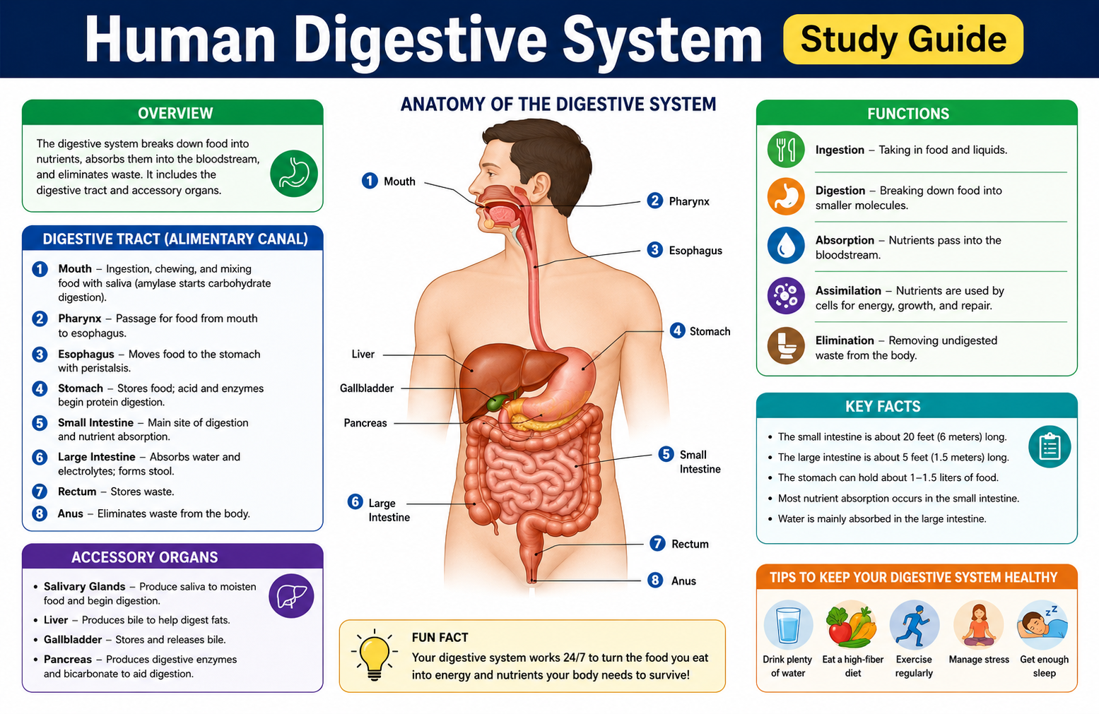
The human digestive system—also called the gastrointestinal (GI) system—is the series of organs responsible for breaking down food into nutrients, absorbing those nutrients into the bloodstream, and eliminating waste materials.
It consists of two main components:
1. The alimentary canal (GI tract) – A continuous, muscular tube approximately 9 meters (about 30 feet) long in adults, extending from the mouth to the anus. It includes: mouth → pharynx → esophagus → stomach → small intestine → large intestine → rectum → anus.
2. Accessory digestive organs – Structures that are not part of the main tube but produce secretions essential for digestion: salivary glands, liver, gallbladder, and pancreas.
The digestive system works through a combination of mechanical processes (physical breakdown through chewing and churning) and chemical processes (enzymatic breakdown of complex molecules into simpler ones that can be absorbed).
A useful analogy: the digestive system is like an industrial food processing facility. Food enters as complex, unusable raw material and exits the other end either as usable nutrients (absorbed into the bloodstream) or as waste (eliminated through defecation). Each section of the GI tract is a specialized processing station with its own specific function.
Why the Digestive System Is Important
Every cell in your body needs energy and building materials. The digestive system is the mechanism that acquires both. Without digestion:
- Glucose couldn’t reach cells for cellular respiration to produce ATP
- Amino acids couldn’t reach tissues for protein synthesis
- Fatty acids couldn’t support membrane structure, hormone production, or fat-soluble vitamin absorption
- Minerals like calcium, iron, and phosphorus couldn’t reach bones, blood, or enzymes
- Water couldn’t be efficiently recovered from ingested material
Beyond energy and nutrients, the digestive system also serves as a critical immune barrier. The gut-associated lymphoid tissue (GALT)—including Peyer’s patches in the small intestine—is one of the largest immune organs in the body, providing surveillance against pathogens that enter through food.
The gut microbiome—the community of trillions of bacteria living in your large intestine—influences not just digestion but also immune function, mental health (via the gut-brain axis), and protection against pathogens.
Functions of the Digestive System
| Function | Description |
|---|---|
| Ingestion | Taking food into the body through the mouth |
| Propulsion | Moving food along the GI tract through swallowing and peristalsis |
| Mechanical digestion | Physical breakdown of food into smaller pieces |
| Chemical digestion | Enzymatic breakdown of large molecules into absorbable monomers |
| Absorption | Transfer of digested nutrients into blood or lymph |
| Assimilation | Incorporation of absorbed nutrients into body cells |
| Elimination | Removal of indigestible waste as feces |
| Immune function | GALT provides immune surveillance and defense |
| Endocrine function | Produces hormones that coordinate digestion |
History of the Study of the Digestive System
The scientific study of digestion has a long and fascinating history, marked by experiments that were sometimes creative, sometimes dramatic, and occasionally stomach-turning.
Ancient and Medieval Knowledge
Ancient Greek physicians understood the basic concept of food processing in the body, though their explanations were tied to humoral theory rather than chemistry. Galen (2nd century CE) described digestive organs based on animal dissections and believed the liver was the center of nutrition.
William Beaumont and Alexis St. Martin (1820s–1830s)
The most famous digestive physiology experiments came from an unlikely partnership. Alexis St. Martin, a fur trapper, was shot in the stomach in 1822 and survived—but with a permanent gastric fistula (an opening from his stomach through his abdominal wall). Army surgeon William Beaumont used this remarkable circumstance to conduct over 200 experiments on gastric digestion, observing directly how different foods were digested, measuring gastric acid production, and demonstrating that gastric juice was chemically active. His findings, published in 1833, established the foundation of gastric physiology.
Ivan Pavlov (late 1800s–early 1900s)
Russian physiologist Ivan Pavlov won the Nobel Prize in 1904 for his work on digestive physiology—particularly his studies of how nervous system signals control digestive secretions. His work on conditioned reflexes began with his observation that dogs salivated not just at food but at anticipatory stimuli—a discovery that revealed the neural control of digestion.
20th Century Advances
The discovery of digestive hormones (secretin in 1902 by Bayliss and Starling, gastrin by Edkins in 1905) revealed that digestion is regulated not just by nerves but by chemical signals. Subsequent decades brought understanding of enzyme mechanisms, gut microbiome function, and the molecular basis of digestive disorders.
Major Organs of the Human Digestive System
Mouth
The mouth (oral cavity) is where digestion begins. It serves as the entry point for food and initiates both mechanical and chemical digestion simultaneously.
Key structures:
- Teeth – Mechanically break food into smaller pieces
- Tongue – Manipulates food, detects taste, and forms the bolus
- Salivary glands – Secrete saliva containing digestive enzymes
The palate (hard and soft) forms the roof of the mouth. The uvula (that small dangling structure at the back of your throat) helps prevent food from entering the nasal cavity during swallowing.
Teeth
Adult humans have 32 teeth designed for different mechanical functions:
- Incisors (8) – Flat, sharp edges for cutting food
- Canines (4) – Pointed; tear and grip food
- Premolars (8) – Crush and grind food
- Molars (12, including wisdom teeth) – Heavy grinding and crushing
The efficiency of chewing directly affects how well the rest of digestion proceeds—poorly chewed food reaches the stomach in larger pieces, requiring more work from gastric mechanical digestion.
Tongue
The tongue is a muscular organ covered with taste buds (papillae) that detect five basic tastes: sweet, salty, sour, bitter, and umami (savory). Beyond taste detection, the tongue:
- Manipulates food during chewing to ensure thorough mechanical processing
- Mixes food with saliva
- Forms the food into a bolus (rounded mass) for swallowing
- Initiates the swallowing reflex
Salivary Glands
Three pairs of salivary glands produce approximately 1–1.5 liters of saliva per day:
- Parotid glands – Largest; located near the ears; produce watery, enzyme-rich saliva
- Submandibular glands – Located under the jaw; produce mixed (serous and mucous) saliva
- Sublingual glands – Located under the tongue; produce mainly mucous saliva
Functions of saliva:
- Moistens food to aid chewing and swallowing
- Contains salivary amylase – begins starch digestion
- Contains lysozyme – antibacterial enzyme
- Contains mucin – lubricates the bolus
- Maintains oral pH and protects teeth from acid
Pharynx
The pharynx is the common passageway for both food and air—a muscular funnel at the back of the mouth and nasal cavity. During swallowing, the epiglottis (a cartilage flap) covers the entrance to the trachea (airway), directing food into the esophagus and preventing choking. The swallowing reflex is involuntary—once initiated, it proceeds automatically.
Esophagus
The esophagus is a muscular tube approximately 25 cm long connecting the pharynx to the stomach. It has no digestive function—its sole role is propulsion.
Food moves through the esophagus via peristalsis—coordinated waves of muscular contraction and relaxation that push the bolus downward. Peristalsis is so effective that you can swallow food while standing on your head—gravity is not required.
At the base of the esophagus is the lower esophageal sphincter (LES) (also called the cardiac sphincter), which relaxes to allow food into the stomach and then closes to prevent stomach acid from refluxing into the esophagus. When the LES doesn’t close properly, the result is acid reflux (GERD).
Stomach
The stomach is a J-shaped, muscular organ in the upper left abdomen that serves as both a storage vessel and a processing chamber. It holds 1–4 liters of material and can stretch considerably after a large meal.
Regions of the stomach:
- Cardia – Connects to the esophagus
- Fundus – Upper region; stores swallowed air and liquid
- Body (corpus) – Main region; primary site of gastric digestion
- Pylorus – Lower region; connects to duodenum via the pyloric sphincter
Stomach wall layers:
The stomach has an additional muscle layer compared to other GI organs—three layers of smooth muscle (longitudinal, circular, and oblique) that enable the churning motion creating chyme (the semi-liquid mixture of partially digested food and gastric juice).
Gastric glands in the stomach lining produce:
- Pepsinogen (converted to pepsin by HCl) – protein digestion
- Hydrochloric acid (HCl) – creates acidic environment (pH 1.5–3.5); kills pathogens; activates pepsin
- Intrinsic factor – essential for vitamin B12 absorption
- Mucus – protects stomach lining from self-digestion
The stomach typically empties into the small intestine over 2–4 hours, regulated by the pyloric sphincter.
Small Intestine
The small intestine is approximately 6–7 meters long—the longest part of the digestive tract—and the primary site of chemical digestion and nutrient absorption. Its inner surface is dramatically amplified by three structural adaptations:
- Circular folds (plicae circulares) – Large folds of mucosa and submucosa
- Villi – Finger-like projections on the folds
- Microvilli (brush border) – Tiny projections on each villus cell
Together, these increase the absorptive surface area to approximately 250 square meters—the size of a tennis court—from a tube that’s only about 2.5 cm wide.
Each villus contains:
- Capillaries – absorb water-soluble nutrients (glucose, amino acids, water-soluble vitamins, minerals) into the bloodstream
- Lacteals – lymphatic vessels that absorb fat-soluble nutrients (fatty acids, fat-soluble vitamins, glycerol) into the lymphatic system
Duodenum
The first 25 cm of the small intestine. Despite being the shortest section, the duodenum is the most biochemically active—it’s where bile (from the liver/gallbladder) and pancreatic juice (from the pancreas) are delivered via the common bile duct and pancreatic duct at the sphincter of Oddi. Most chemical digestion is completed in the duodenum.
Jejunum
The middle section (~2.5 meters). The jejunum is the primary site of nutrient absorption—its villi are particularly tall and densely packed with transport proteins. Most carbohydrates, proteins, water-soluble vitamins, and minerals are absorbed here.
Ileum
The final and longest section (~3.5 meters). The ileum absorbs remaining nutrients, particularly vitamin B12 (bound to intrinsic factor) and bile salts (recycled back to the liver—enterohepatic circulation). The ileum connects to the large intestine at the ileocecal valve.
Large Intestine (Colon)
The large intestine is approximately 1.5 meters long and 6–7 cm in diameter—shorter but wider than the small intestine. It receives the unabsorbed material from the ileum and performs several important functions:
- Water absorption – Recovers approximately 1–1.5 liters of water per day from liquid chyme, converting it to semi-solid feces
- Electrolyte absorption – Absorbs sodium, potassium, and chloride
- Mucus secretion – Lubricates feces for movement
- Vitamin production – Gut bacteria produce vitamin K and some B vitamins
- Feces formation – Compacts undigestible material into feces
Regions of the large intestine:
- Cecum (with appendix)
- Ascending colon
- Transverse colon
- Descending colon
- Sigmoid colon
Rectum
The rectum is approximately 15 cm long and serves as the temporary storage site for feces. When feces accumulate and stretch the rectal walls, sensory nerves signal the urge to defecate. The rectum connects to the anus via the anal canal.
Anus
The anus is the terminal opening of the digestive tract through which feces are eliminated. It has two sphincters:
- Internal anal sphincter – Smooth muscle; involuntary control
- External anal sphincter – Skeletal muscle; voluntary control (allows conscious control of defecation)
Accessory Digestive Organs
These organs don’t form part of the alimentary canal but produce essential secretions that are delivered into the GI tract.
Liver
The liver is the largest internal organ (approximately 1.5 kg) and one of the most metabolically complex organs in the body. Its digestive function centers on producing bile.
Digestive functions of the liver:
- Bile production – Produces 500–1000 mL of bile per day; bile contains bile salts that emulsify fats, making them accessible to lipase
- Nutrient processing – After absorption, nutrients from the small intestine travel to the liver via the hepatic portal vein for processing: glucose is stored as glycogen or converted to fat; amino acids are processed; toxins are detoxified
- Vitamin storage – Stores fat-soluble vitamins (A, D, E, K) and vitamin B12
Gallbladder
The gallbladder is a small, pear-shaped organ tucked under the liver. It concentrates and stores bile produced by the liver—up to 10-fold concentration. When fatty food enters the duodenum, the hormone cholecystokinin (CCK) triggers the gallbladder to contract, releasing concentrated bile through the common bile duct into the duodenum.
Gallstones form when bile becomes supersaturated with cholesterol or bilirubin, crystallizing into stones that can block the bile duct—causing intense pain (biliary colic).
Pancreas
The pancreas is both an endocrine organ (producing insulin and glucagon to regulate blood sugar) and an exocrine organ (producing pancreatic juice for digestion).
Pancreatic juice contains:
- Pancreatic amylase – continues starch digestion
- Pancreatic lipase – primary fat-digesting enzyme
- Trypsin, chymotrypsin, elastase – protein-digesting enzymes (produced as inactive precursors to prevent self-digestion)
- Pancreatic nucleases – digest nucleic acids (DNA and RNA)
- Sodium bicarbonate (NaHCO₃) – neutralizes acidic chyme entering the duodenum from the stomach, raising pH to ~7–8 for optimal enzyme function
Complete Process of Digestion
Ingestion
Taking food into the body through the mouth. The sight, smell, and taste of food trigger anticipatory secretions (cephalic phase of digestion)—saliva production increases, gastric acid secretion begins. This is Pavlov’s conditioned reflex in action—your mouth actually does water before food arrives.
Mechanical Digestion
Physical breakdown of food into smaller pieces without chemical change. Increases surface area for enzyme action.
Where it occurs:
- Mouth – Chewing (mastication) by teeth
- Stomach – Churning by muscular walls creating chyme
- Small intestine – Segmentation contractions mix chyme with digestive juices
Chemical Digestion
Enzymatic breakdown of large molecules into monomers:
- Polysaccharides → monosaccharides (glucose, fructose, galactose)
- Proteins → amino acids
- Fats → fatty acids + glycerol
- Nucleic acids → nucleotides
Occurs throughout the GI tract—saliva in the mouth, gastric juice in the stomach, and bile + pancreatic juice + intestinal juice in the small intestine.
Absorption
Transfer of digested nutrients from the small intestine (primarily) into the blood or lymph for distribution throughout the body. Most absorption occurs in the jejunum, with some in the duodenum and ileum.
Assimilation
The use of absorbed nutrients by body cells—glucose in cellular respiration, amino acids in protein synthesis, fatty acids in membrane construction and energy storage. This is the actual purpose the whole system exists to serve.
Elimination (Defecation)
Removal of indigestible waste (fiber, dead cells, bacteria, water) as feces through the anus. The large intestine takes approximately 12–72 hours to complete this final stage.
Digestive Enzymes Explained
| Enzyme | Produced By | Location of Action | Substrate | Product |
|---|---|---|---|---|
| Salivary amylase | Salivary glands | Mouth | Starch (polysaccharide) | Maltose |
| Pepsin | Stomach (chief cells) | Stomach | Proteins | Peptides |
| Gastric lipase | Stomach | Stomach | Fats | Fatty acids + glycerol (partial) |
| Pancreatic amylase | Pancreas | Small intestine | Starch | Maltose |
| Pancreatic lipase | Pancreas | Small intestine | Fats (emulsified) | Fatty acids + glycerol |
| Trypsin | Pancreas | Small intestine | Proteins/peptides | Smaller peptides |
| Chymotrypsin | Pancreas | Small intestine | Proteins/peptides | Smaller peptides |
| Maltase | Small intestine | Small intestine | Maltose | Glucose + Glucose |
| Lactase | Small intestine | Small intestine | Lactose | Glucose + Galactose |
| Sucrase | Small intestine | Small intestine | Sucrose | Glucose + Fructose |
| Peptidases | Small intestine | Small intestine | Peptides | Amino acids |
Amylase
Breaks starch (a polysaccharide) into maltose (a disaccharide). Salivary amylase begins starch digestion in the mouth—if you chew a piece of bread long enough, it starts tasting sweet as maltose accumulates. Pancreatic amylase continues this work in the small intestine.
Pepsin
The primary protein-digesting enzyme of the stomach. It’s produced in an inactive form called pepsinogen by chief cells—if it were produced in active form, it would digest the stomach wall itself. Pepsinogen is activated to pepsin by the acidic environment (HCl) of the stomach.
Trypsin
A powerful protease produced by the pancreas as inactive trypsinogen. Activated in the duodenum by the enzyme enterokinase (produced by duodenal cells). Trypsin then activates other pancreatic enzyme precursors (chymotrypsinogen → chymotrypsin; proelastase → elastase)—a cascading activation sequence.
Lipase
Pancreatic lipase is the primary fat-digesting enzyme. It breaks triglycerides into fatty acids and monoglycerides. Works only on emulsified fat—bile salts from the liver must first break large fat globules into tiny droplets (emulsification) to expose sufficient surface area for lipase to work.
Maltase, Lactase, Sucrase
These brush border enzymes are produced by intestinal cells and embedded in the microvilli surface. They complete carbohydrate digestion by breaking disaccharides into monosaccharides ready for absorption. Lactase deficiency is the basis of lactose intolerance—without this enzyme, lactose reaches the large intestine undigested, where bacteria ferment it, producing gas and osmotic diarrhea.
Digestive Juices and Their Functions
Saliva
- Volume: ~1–1.5 liters/day
- pH: 6.5–7.5 (slightly acidic to neutral)
- Components: Water, mucin, salivary amylase, lysozyme, immunoglobulin A, bicarbonate
- Functions: Moisten food; begin starch digestion; lubricate bolus; antibacterial; protect teeth
Gastric Juice
- Volume: ~2–3 liters/day
- pH: 1.5–3.5 (strongly acidic)
- Components: HCl, pepsinogen, intrinsic factor, gastric lipase, mucus
- Functions: Kill pathogens; denature food proteins; activate pepsin; begin protein digestion; enable B12 absorption (intrinsic factor)
Bile
- Volume: ~500–1000 mL/day
- pH: 7.6–8.6 (alkaline)
- Components: Bile salts, bilirubin (bile pigment from hemoglobin breakdown), cholesterol, phospholipids, water
- Functions: Emulsify fats (the crucial function—increases fat surface area for lipase); excrete bilirubin waste; alkalinize duodenal contents
Pancreatic Juice
- Volume: ~1.5 liters/day
- pH: 8–8.3 (alkaline)
- Components: Sodium bicarbonate, pancreatic amylase, lipase, trypsinogen, chymotrypsinogen, nucleases
- Functions: Neutralize acidic chyme; complete digestion of all major food groups
Intestinal Juice (Succus Entericus)
- Volume: ~1–2 liters/day
- pH: ~7.6
- Components: Water, mucus, brush border enzymes (maltase, lactase, sucrase, peptidases)
- Functions: Final stages of carbohydrate and protein digestion
How Nutrients Are Absorbed
Carbohydrates
Polysaccharides → disaccharides (by amylase) → monosaccharides (glucose, galactose, fructose by brush border enzymes).
Glucose and galactose are absorbed by active transport (secondary active transport via sodium-glucose transporter SGLT1). Fructose enters by facilitated diffusion. Once inside intestinal cells, all monosaccharides exit via the GLUT2 transporter into capillaries of the villi, traveling to the liver via the hepatic portal vein.
Proteins
Proteins → peptides (by pepsin, trypsin, chymotrypsin) → amino acids and small peptides (by brush border peptidases).
Amino acids are absorbed by active transport via specific amino acid transporters. Small peptides (di- and tripeptides) enter intestinal cells directly and are hydrolyzed inside the cell. Amino acids then diffuse into capillaries and travel to the liver for processing.
Fats
Triglycerides → emulsified fat droplets (by bile salts) → fatty acids + monoglycerides (by pancreatic lipase).
These products form micelles—tiny aggregates with bile salts—that transport fatty acids and monoglycerides to the intestinal cell surface. Inside intestinal cells, fatty acids and monoglycerides are reassembled into triglycerides, packaged with proteins into chylomicrons, and released into lacteals (lymphatic vessels). Fat-soluble vitamins (A, D, E, K) follow the same pathway.
Vitamins
- Water-soluble vitamins (B vitamins, C): Absorbed primarily in the jejunum by active transport or facilitated diffusion; directly into capillaries
- Fat-soluble vitamins (A, D, E, K): Absorbed with dietary fat via micelles and chylomicrons into lacteals
- Vitamin B12: Unique—must bind to intrinsic factor (produced by stomach) and is absorbed in the ileum by specific receptors
Minerals
- Iron: Absorbed in duodenum; absorption enhanced by vitamin C (forms soluble complex) and inhibited by phytates (in whole grains)
- Calcium: Absorbed in duodenum and jejunum; requires vitamin D for active transport; absorption increases when dietary calcium is low
- Sodium: Absorbed throughout small intestine; drives co-transport of glucose and amino acids
Water
Approximately 9 liters of water enter the GI tract daily (2 liters from diet; 7 liters from digestive secretions). The small intestine absorbs approximately 7.5 liters; the large intestine absorbs ~1.5 liters; only ~100–150 mL is lost in feces. This efficient water recovery is critical—failure of the large intestine to absorb water (as in diarrhea) leads rapidly to dehydration.
Human Digestive System Diagram Explained
A complete diagram of the human digestive system shows:
MOUTH
  ↓ (Pharynx)
ESOPHAGUS (25 cm)
  ↓ (Lower esophageal sphincter)
STOMACH (J-shaped; 1–4 L capacity)
  ↓ (Pyloric sphincter)
SMALL INTESTINE (6–7 m)
  ├── Duodenum (25 cm) → receives bile + pancreatic juice
  ├── Jejunum (2.5 m) → primary absorption
  └── Ileum (3.5 m) → B12 + bile salt absorption
  ↓ (Ileocecal valve)
LARGE INTESTINE (1.5 m)
  ├── Cecum + Appendix
  ├── Ascending colon
  ├── Transverse colon
  ├── Descending colon
  └── Sigmoid colon
  ↓
RECTUM (15 cm)
  ↓
ANUS
Accessory organs (connected via ducts):
- Salivary glands → mouth
- Liver → produces bile → gallbladder stores → duodenum
- Pancreas → produces pancreatic juice → duodenum
Journey of Food Through the Digestive Tract
Let’s follow a meal—say, a cheese sandwich—through the complete digestive journey:
- Mouth (seconds to minutes): Teeth mechanically break bread, cheese, and any fillings into smaller pieces. Salivary amylase begins breaking bread starch into maltose. Tongue forms the mixture into a bolus. Swallowing occurs—epiglottis covers trachea.
- Esophagus (~10 seconds): Peristalsis propels the bolus to the stomach.
- Stomach (2–4 hours): HCl denatures proteins in the cheese and any meat. Pepsin begins protein digestion. Muscular churning creates chyme. Gastric lipase begins fat breakdown. The acidic chyme is released into the duodenum in small, regulated portions through the pyloric sphincter.
- Duodenum (minutes to hours): Bile emulsifies fats. Pancreatic juice neutralizes acidity and delivers amylase (starch), lipase (fats), and proteases (proteins). Most chemical digestion is completed here.
- Jejunum (1–4 hours): Brush border enzymes complete carbohydrate and protein digestion. Glucose, amino acids, fatty acids, vitamins, and minerals are absorbed across the highly folded, villus-covered wall into capillaries and lacteals.
- Ileum (1–4 hours): Remaining absorption—vitamin B12, bile salts. Unabsorbed material passes to the large intestine.
- Large intestine (10–59 hours): Water is absorbed. Gut bacteria ferment undigested fiber, producing short-chain fatty acids (which nourish colon cells) and gases. Feces form.
- Rectum: Feces stored until defecation reflex is triggered.
- Anus: Feces eliminated.
Total transit time: Approximately 24–72 hours in a healthy adult.
Human Digestive System Flowchart
FOOD INTAKE
    ↓
MOUTH → Chewing + Salivary amylase → BOLUS formed
    ↓
ESOPHAGUS → Peristalsis
    ↓
STOMACH → HCl + Pepsin + Churning → CHYME formed
    ↓
DUODENUM → Bile (emulsification) + Pancreatic juice (enzymes) → Chemical digestion completed
    ↓
JEJUNUM → Absorption of: Glucose / Amino acids / Fatty acids / Vitamins / Minerals
    ↓
ILEUM → Absorption of: Vitamin B12 / Bile salts
    ↓
LARGE INTESTINE → Water absorption / Electrolyte absorption / Bacterial fermentation → FECES formed
    ↓
RECTUM → Storage
    ↓
ANUS → Elimination
Mechanical Digestion vs Chemical Digestion
| Feature | Mechanical Digestion | Chemical Digestion |
|---|---|---|
| Definition | Physical breakdown of food into smaller pieces | Enzymatic breakdown of large molecules into monomers |
| Type of change | Physical change (no molecular change) | Chemical change (molecular bonds broken) |
| Organs involved | Mouth, stomach, small intestine | Mouth, stomach, small intestine, pancreas |
| Enzymes used | No enzymes | Yes—amylase, pepsin, lipase, trypsin, etc. |
| Process | Chewing, churning, segmentation | Hydrolysis reactions |
| Purpose | Increase surface area for enzyme action | Break down nutrients into absorbable forms |
| Examples | Chewing bread; stomach churning protein | Amylase breaking starch to maltose |
| End product | Smaller pieces of the same food | Monomers: glucose, amino acids, fatty acids |
Digestion of Carbohydrates
Step 1 (Mouth): Salivary amylase breaks starch into maltose and dextrins. (Brief—stops when food reaches acidic stomach.)
Step 2 (Small intestine): Pancreatic amylase continues starch → maltose/dextrins.
Step 3 (Brush border): Maltase → glucose + glucose; Lactase → glucose + galactose; Sucrase → glucose + fructose.
Absorption: Monosaccharides absorbed into capillaries → hepatic portal vein → liver → bloodstream.
Digestion of Proteins
Step 1 (Stomach): HCl denatures protein structure; pepsin cleaves peptide bonds → short peptides.
Step 2 (Duodenum/Small intestine): Trypsin, chymotrypsin, and elastase further cleave peptides.
Step 3 (Brush border): Peptidases break remaining peptides → amino acids; some di/tripeptides absorbed directly.
Absorption: Amino acids transported into capillaries → hepatic portal vein → liver.
Digestion of Fats
Step 1 (Stomach): Gastric lipase begins minimal fat digestion.
Step 2 (Duodenum): Bile salts emulsify fat globules into tiny droplets (emulsification—not digestion, but essential for it).
Step 3 (Small intestine): Pancreatic lipase breaks triglycerides into fatty acids + monoglycerides.
Step 4 (Micelle formation): Bile salts + fatty acids + monoglycerides form micelles that transport products to intestinal cell surface.
Step 5 (Absorption): Products diffuse into intestinal cells → reassembled into triglycerides → packaged into chylomicrons → released into lacteals → lymphatic system → bloodstream (via thoracic duct into left subclavian vein).
Role of the Gut Microbiome
The gut microbiome is the community of approximately 38 trillion microorganisms—predominantly bacteria—living primarily in the large intestine. Far from being passive passengers, these microorganisms are active participants in digestion and overall health.
Digestive functions:
- Ferment dietary fiber – Produce short-chain fatty acids (SCFAs: butyrate, propionate, acetate) that nourish colonocytes (colon cells) and have anti-inflammatory effects
- Synthesize vitamins – Produce vitamin K and B vitamins (biotin, folate)
- Break down compounds – Metabolize bile acids, plant polyphenols, and drug compounds
- Competitive exclusion – Prevent colonization by pathogenic bacteria
Beyond digestion:
- Immune regulation – 70% of the body’s immune cells are in the gut; microbiome shapes immune responses
- Gut-brain axis – Gut bacteria communicate with the brain via the vagus nerve and produce neurotransmitters (including 95% of the body’s serotonin)
- Mental health – Alterations in gut microbiome composition are associated with depression, anxiety, and autism spectrum conditions
Factors affecting microbiome health: Diet (particularly fiber intake), antibiotics, stress, age, birth method (vaginal delivery vs C-section), and breastfeeding all influence microbiome composition.
Digestive Hormones
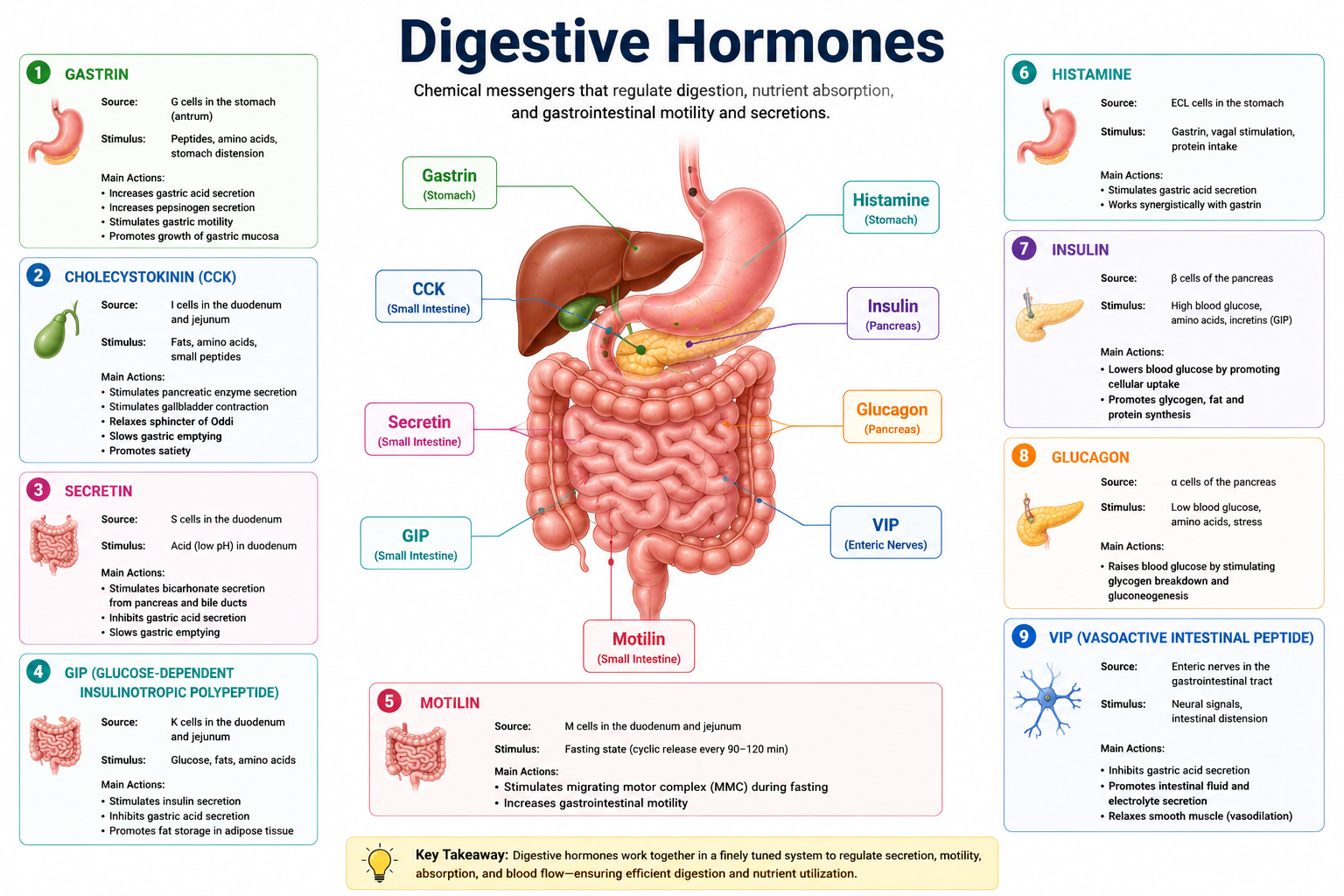
Gastrin
- Produced by: G cells in the stomach antrum
- Trigger: Protein in stomach; distension; vagal nerve signals
- Function: Stimulates gastric acid (HCl) secretion by parietal cells; promotes gastric motility; stimulates pepsinogen secretion
- Clinical note: Gastrinoma (Zollinger-Ellison syndrome) is a tumor that produces excess gastrin, causing severe peptic ulcers
Secretin
- Produced by: S cells in the duodenum
- Trigger: Acidic chyme entering the duodenum
- Function: Stimulates pancreas to secrete bicarbonate (neutralizing duodenal acid); inhibits gastric acid secretion and motility; stimulates bile secretion
- Historical note: Secretin (discovered 1902) was the first hormone ever identified—its discovery by Bayliss and Starling introduced the concept of chemical messengers in the body
Cholecystokinin (CCK)
- Produced by: I cells in the duodenum and jejunum
- Trigger: Fat and protein in the small intestine
- Function: Stimulates gallbladder contraction (bile release); stimulates pancreatic enzyme secretion; slows gastric emptying; signals satiety to the brain
- Clinical relevance: CCK’s satiety signaling explains why fat-containing meals keep you full longer than carbohydrate-only meals
Common Digestive Disorders
Acid Reflux (GERD)
Cause: Lower esophageal sphincter doesn’t close properly, allowing stomach acid to enter the esophagus.
Symptoms: Heartburn, regurgitation, chest discomfort.
Treatment: Lifestyle changes (avoid trigger foods, elevate head when sleeping), antacids, H2 blockers, proton pump inhibitors (PPIs).
Gastritis
Cause: Inflammation of the stomach lining, often from H. pylori infection, NSAIDs, or alcohol.
Symptoms: Upper abdominal pain, nausea, bloating.
Treatment: Antibiotics (for H. pylori), acid-reducing medications, avoiding irritants.
Peptic Ulcer
Cause: Erosion of the mucosal lining of the stomach or duodenum, most commonly from H. pylori or NSAID use.
Symptoms: Burning stomach pain (often relieved by food in gastric ulcers; worsened by food in duodenal ulcers), nausea.
Treatment: Antibiotics for H. pylori, PPIs, avoid NSAIDs.
Constipation
Cause: Slow transit time, inadequate fiber/water intake, sedentary lifestyle, medications.
Symptoms: Infrequent bowel movements (<3/week), hard/dry stools, straining.
Treatment: Increased fiber, water, exercise; laxatives if needed.
Diarrhea
Cause: Infection (viral, bacterial, parasitic), food intolerance, IBS, inflammatory bowel disease, medications.
Symptoms: Loose, watery stools more than 3 times per day.
Treatment: Rehydration (critical—especially in children), treating underlying cause.
Irritable Bowel Syndrome (IBS)
Cause: Not fully understood; involves abnormal gut motility, visceral hypersensitivity, gut-brain axis disruption, microbiome changes.
Symptoms: Abdominal pain, bloating, alternating constipation and diarrhea.
Treatment: Dietary modification (low-FODMAP diet), stress management, probiotics, medications.
Crohn’s Disease
Cause: Chronic inflammatory bowel disease; immune-mediated; can affect any part of the GI tract from mouth to anus; transmural inflammation.
Symptoms: Abdominal pain, diarrhea, weight loss, fatigue, perianal disease.
Treatment: Anti-inflammatory medications, immunosuppressants, biologics, surgery.
Ulcerative Colitis
Cause: Chronic inflammatory bowel disease; limited to the colon and rectum; mucosal inflammation.
Symptoms: Bloody diarrhea, abdominal cramping, urgency.
Treatment: Aminosalicylates, steroids, immunosuppressants, biologics; surgery (colectomy) can be curative.
Gallstones
Cause: Crystallization of cholesterol or bilirubin in bile; risk factors include obesity, female sex, rapid weight loss, certain medications.
Symptoms: Often asymptomatic; biliary colic (severe right upper quadrant pain) if stones obstruct bile duct.
Treatment: Surgical removal of gallbladder (cholecystectomy); bile acid medications for small stones.
Lactose Intolerance
Cause: Deficiency of lactase enzyme; common in adults, particularly in East Asian, African, and Native American populations.
Symptoms: Bloating, gas, diarrhea after consuming dairy products.
Treatment: Limit lactose intake, use lactase supplements, choose lactose-free products.
Celiac Disease
Cause: Autoimmune reaction to gluten (protein in wheat, barley, rye); immune attack damages villi of small intestine → malabsorption.
Symptoms: Diarrhea, bloating, weight loss, nutritional deficiencies, fatigue.
Treatment: Strict lifelong gluten-free diet—the only effective treatment currently.
Tips for Maintaining a Healthy Digestive System
- Eat adequate dietary fiber (25–38 grams/day) – supports bowel regularity and microbiome health
- Stay well hydrated – water is essential for stool formation and mucosal health
- Exercise regularly – physical activity promotes gut motility
- Chew food thoroughly – reduces the digestive burden on the stomach and intestines
- Eat at regular intervals – helps regulate digestive hormone cycles
- Limit processed foods and excessive alcohol – both damage the gut lining and disrupt the microbiome
- Manage stress – the gut-brain axis means chronic stress directly impairs digestion
- Don’t ignore the urge to defecate – delaying consistently can lead to constipation
- Use antibiotics only when necessary – broad-spectrum antibiotics disrupt the microbiome significantly
- Include probiotic and prebiotic foods – yogurt, kefir, sauerkraut, garlic, onion support microbiome health
Foods That Support Digestion
- Fiber-rich foods: Oats, legumes, vegetables, fruits—support bowel regularity and microbiome
- Probiotic foods: Yogurt, kefir, kimchi, sauerkraut, miso—provide beneficial bacteria
- Prebiotic foods: Garlic, onions, bananas, asparagus—feed beneficial bacteria
- Water-rich foods: Cucumber, watermelon, celery—support hydration and digestion
- Ginger: Has anti-nausea and pro-motility effects
- Papaya: Contains papain—a natural protein-digesting enzyme
Foods That May Harm Digestion
- Highly processed foods – Low fiber; high in additives that may disrupt microbiome
- Excessive alcohol – Damages gastric and intestinal lining; disrupts microbiome
- Fried/fatty foods – Slow gastric emptying; trigger acid reflux and gallbladder symptoms
- Spicy foods – Can irritate the esophageal and gastric lining in susceptible individuals
- Dairy (for lactose intolerant individuals) – Causes gas, bloating, diarrhea
- Excessive caffeine – Can stimulate excessive gastric acid secretion
Digestive System in Children
Children’s digestive systems are anatomically similar to adults but functionally immature at birth. Newborns:
- Have lower gastric acid production (gradually increases over the first months)
- Begin life entirely dependent on breast milk or formula—easily digestible complete nutrition
- Lack the enzyme amylase in sufficient quantities until approximately 6 months (which is why starchy solid foods are introduced around this time)
- Have a highly permeable intestinal lining (“leaky gut”) in early infancy—important for absorbing maternal antibodies from breast milk but also means greater susceptibility to allergen sensitization
Common digestive issues in children include colic, gastroesophageal reflux, constipation, and food allergies. The microbiome is established in the first years of life—shaped by birth method, breastfeeding, and early diet—with lasting effects on health.
Digestive System Changes with Aging
As people age, digestive function changes in several important ways:
- Reduced saliva production – Dry mouth is common; impairs initial digestion and swallowing
- Decreased gastric acid secretion – Impairs B12 absorption, iron absorption, and protein digestion; increases susceptibility to foodborne illness
- Slower peristalsis – Contributes to constipation—the most common GI complaint in older adults
- Reduced enzyme production – Particularly lactase, explaining increasing lactose intolerance with age
- Decreased liver function – Slower drug metabolism; reduced bile acid production
- Altered microbiome – Reduced diversity; decrease in beneficial Bifidobacterium species
- Increased risk of diverticulosis – Outpouchings in the colon wall, common after age 60
Interesting Facts About the Digestive System
Important Facts Box:
- The small intestine, though narrower, is actually much longer than the large intestine—approximately 6–7 meters vs 1.5 meters
- If the small intestine’s surface were flattened, it would cover a tennis court (~250 m²)
- The stomach produces a new mucus lining every two weeks to protect against self-digestion
- About 1.3 kg of bacteria live in your large intestine—more bacteria than human cells in your body
- The esophagus doesn’t require gravity—peristalsis allows swallowing upside down
- You produce approximately 7 liters of digestive secretions per day
- The gut contains approximately 100 million neurons—more than the spinal cord
- The appendix, once thought vestigial, is now thought to serve as a “safe house” for beneficial gut bacteria after infection
- 95% of the body’s serotonin is produced in the gut, not the brain
- Gastric acid is strong enough to dissolve zinc metal
Common Biology Terms Every Student Should Know
| Term | Definition |
|---|---|
| Peristalsis | Wave-like muscular contractions propelling food through the GI tract |
| Bolus | Rounded mass of food formed in the mouth before swallowing |
| Chyme | Semi-liquid mixture of partially digested food and gastric juice in the stomach |
| Emulsification | Breaking large fat globules into small droplets (by bile salts) |
| Hydrolysis | Chemical digestion—breaking molecules using water |
| Villi | Finger-like projections on the small intestinal lining increasing surface area |
| Microvilli | Tiny projections on villus cells forming the brush border |
| Lacteals | Lymphatic vessels within villi that absorb fats and fat-soluble vitamins |
| Sphincter | Muscular valve controlling movement of material between GI segments |
| Intrinsic factor | Glycoprotein produced by stomach; essential for vitamin B12 absorption |
| Enterohepatic circulation | Recycling of bile salts from ileum back to liver |
| Chylomicrons | Lipoprotein particles carrying fat from intestinal cells into lymphatics |
| GALT | Gut-associated lymphoid tissue; immune surveillance in the gut |
| Rugae | Folds in the stomach lining that allow expansion |
| Segmentation | Rhythmic contractions in the small intestine mixing chyme |
Common Mistakes Students Make
1. Confusing the small and large intestines by size
The small intestine is narrow (about 2.5 cm diameter) but very long (6–7 m). The large intestine is wide (6–7 cm) but short (1.5 m). “Small” and “large” refer to diameter, not length. This is one of the most reliable trick question setups in anatomy exams.
2. Saying bile digests fats
Bile does NOT chemically digest fats. It emulsifies them—breaks fat globules into tiny droplets, increasing surface area. The actual chemical digestion is done by lipase (a pancreatic enzyme). Bile has no enzymatic activity.
3. Forgetting that pepsin is produced as pepsinogen
Pepsin is the active enzyme, but it’s produced as the inactive precursor pepsinogen. It’s activated by HCl in the stomach. If an exam asks where pepsin comes from, the technically correct answer involves pepsinogen from chief cells, not pepsin directly.
4. Confusing absorption sites
Most absorption occurs in the jejunum. Vitamin B12 is specifically absorbed in the ileum. Iron and calcium are primarily absorbed in the duodenum. These specific sites are frequently tested.
5. Describing the liver as only a digestive organ
The liver’s digestive function (bile production) is just one of over 500 known functions. It also detoxifies substances, produces plasma proteins, regulates blood glucose, stores glycogen and fat-soluble vitamins, and processes absorbed nutrients from the portal blood.
Exam Tips
Exam Strategy Box:
- Learn the GI tract in order from mouth to anus—every organ, every sphincter between them
- For enzyme questions: Know the enzyme name, where it’s produced, where it acts, what it breaks down, and what it produces
- For absorption questions: Know whether water-soluble nutrients go to capillaries or fat-soluble nutrients go to lacteals
- For hormones: Know the trigger (what stimulates secretion) and the target organ and effect
- Diagram practice: Be able to draw a labeled diagram of the digestive system from memory
- Disorder questions: Know the affected organ/mechanism—not just the name and symptoms
- Comparison questions: Know mechanical vs chemical digestion cold—this appears in virtually every GI exam
Memory Tricks to Remember Digestive Organs
Remembering the order of organs:
“My Pharynx Easily Swallows Slimy Digestive Junk Like Rotting Anything”
→ Mouth, Pharynx, Esophagus, Stomach, Small intestine (Duodenum, Jejunum, Ileum), Large intestine, Rectum, Anus
Remembering the three parts of the small intestine in order:
“Don’t Jump In” → Duodenum, Jejunum, Ileum
Remembering the regions of the large intestine:
“Cats And Tigers Don’t Shy Away” → Cecum, Ascending, Transverse, Descending, Sigmoid, Anus
Remembering bile vs lipase:
Bile Emulsifies; Lipase Digests (BELT → Bile Emulsifies Lipid To droplets)
Remembering fat-soluble vitamins (go via lacteals):
“Fat DEAK” → Fat-soluble: D, E, A, K
Human Digestive System Practice Questions
20 Multiple Choice Questions with Answers
- Where does chemical digestion of starch begin?
- A) Stomach
- B) Duodenum
- C) Mouth ✓
- D) Jejunum
- Which enzyme is responsible for activating other pancreatic enzymes?
- A) Lipase
- B) Amylase
- C) Pepsin
- D) Trypsin ✓
- The primary function of bile is to:
- A) Digest proteins
- B) Neutralize stomach acid
- C) Emulsify fats ✓
- D) Absorb glucose
- Vitamin B12 is absorbed in which part of the small intestine?
- A) Duodenum
- B) Jejunum
- C) Ileum ✓
- D) Large intestine
- Which structure prevents food from entering the trachea during swallowing?
- A) Uvula
- B) Epiglottis ✓
- C) Soft palate
- D) Larynx
- Where does most nutrient absorption occur?
- A) Duodenum
- B) Stomach
- C) Jejunum ✓
- D) Large intestine
- Which hormone triggers the gallbladder to release bile?
- A) Gastrin
- B) Secretin
- C) Insulin
- D) Cholecystokinin (CCK) ✓
- Fat-soluble vitamins are absorbed into:
- A) Capillaries in the villi
- B) The portal vein
- C) Lacteals ✓
- D) The hepatic artery
- The stomach lining is protected from self-digestion by:
- A) Pepsinogen
- B) Mucus ✓
- C) HCl
- D) Bile
- Which of the following is NOT a function of the large intestine?
- A) Water absorption
- B) Feces formation
- C) Chemical digestion of proteins ✓
- D) Bacterial fermentation of fiber
- Lactose intolerance results from deficiency of:
- A) Amylase
- B) Pepsin
- C) Lactase ✓
- D) Sucrase
- The acidity of gastric juice is maintained at approximately:
- A) pH 6–7
- B) pH 4–5
- C) pH 1.5–3.5 ✓
- D) pH 7–8
- Which accessory organ produces both digestive enzymes and hormones?
- A) Liver
- B) Gallbladder
- C) Salivary glands
- D) Pancreas ✓
- Intrinsic factor, essential for B12 absorption, is produced by:
- A) Chief cells of the stomach
- B) Parietal cells of the stomach ✓
- C) Duodenal cells
- D) Liver cells
- Which of the following describes peristalsis?
- A) Chemical breakdown of food by enzymes
- B) Emulsification of fats by bile
- C) Wave-like muscular contractions propelling food ✓
- D) Absorption of nutrients into the bloodstream
- The sphincter between the stomach and duodenum is called:
- A) Lower esophageal sphincter
- B) Ileocecal valve
- C) Internal anal sphincter
- D) Pyloric sphincter ✓
- Where are chylomicrons formed?
- A) Liver cells
- B) Intestinal epithelial cells ✓
- C) Pancreatic cells
- D) Red blood cells
- Celiac disease damages which intestinal structures?
- A) Rugae
- B) Microvilli only
- C) Villi ✓
- D) Circular folds
- The secretion of pancreatic bicarbonate is triggered by:
- A) CCK
- B) Gastrin
- C) Secretin ✓
- D) Insulin
- How long is the total gastrointestinal tract in adults?
- A) ~3 meters
- B) ~6 meters
- C) ~9 meters ✓
- D) ~12 meters
10 Short Answer Questions
- Describe the role of bile in fat digestion. Is bile an enzyme? Explain why or why not.
- Explain why pancreatic enzymes (trypsin, chymotrypsin) are produced as inactive precursors. What activates them in the small intestine?
- Compare the structure of the small intestine to the large intestine in terms of length, diameter, and absorptive surface adaptations.
- Describe the three stages of gastric secretion (cephalic, gastric, and intestinal phases). What triggers each phase?
- Explain the path taken by dietary fat from ingestion to entry into the lymphatic system. Name all key structures and processes involved.
- What is the enterohepatic circulation of bile salts? Where are bile salts absorbed, and why is this recycling system important?
- Describe the functions of the liver that are relevant to the digestive system. Why is the hepatic portal circulation important?
- Explain how the surface area of the small intestine is increased to maximize absorption. Describe all three structural adaptations.
- What is the gut microbiome and what roles does it play in digestion and overall health?
- Explain the physiological basis of lactose intolerance. Why does it cause the specific symptoms of bloating, gas, and diarrhea?
5 Long Answer Questions
- Describe the complete digestion and absorption of a meal containing carbohydrates, protein, and fat. For each macronutrient, trace the process from ingestion to absorption, naming all enzymes, their locations of action, and the final absorbed products. Explain how fat absorption differs fundamentally from carbohydrate and protein absorption.
- Describe the structure and function of each major organ of the digestive system from mouth to anus. For each organ, describe its anatomical features, the mechanical and/or chemical processes it performs, and the key secretions it produces. Include the roles of the salivary glands, liver, gallbladder, and pancreas as accessory organs.
- Explain the roles of digestive hormones in coordinating the digestive process. For gastrin, secretin, and cholecystokinin: describe the stimulus for secretion, the cells that produce each hormone, the target organs, and the specific effects on digestive processes. Explain how these hormones collectively ensure efficient digestion.
- Discuss ten common digestive disorders, including for each: the organ or system affected, the underlying pathophysiology (cause), the main clinical symptoms, and the primary treatment approach. Organize your answer to show relationships between related conditions where they exist (e.g., GERD and peptic ulcers; Crohn’s disease and ulcerative colitis).
- Describe the structure of the small intestine wall in detail, explaining how it is adapted for maximal absorption. Explain how each of the following nutrients is absorbed: glucose, amino acids, fatty acids, vitamin B12, iron, and calcium. For each, specify the mechanism of transport (active transport, passive diffusion, etc.) and the path taken from intestinal lumen to bloodstream or lymph.
Revision Notes
Quick Revision Summary:
Organs in order: Mouth → Pharynx → Esophagus → Stomach → Small intestine (Duodenum, Jejunum, Ileum) → Large intestine → Rectum → Anus
Accessory organs: Salivary glands, Liver, Gallbladder, Pancreas
Key enzymes: Salivary + Pancreatic amylase (starch), Pepsin (proteins, stomach), Trypsin (proteins, small intestine), Pancreatic lipase (fats), Maltase/Lactase/Sucrase (disaccharides, brush border)
Bile: Produced by liver, stored in gallbladder, released into duodenum, EMULSIFIES fats (not digestion)
Pancreatic juice: Enzymes + bicarbonate; neutralizes acid + digests all macronutrients
Hormones: Gastrin → ↑HCl; Secretin → ↑bicarbonate; CCK → gallbladder contraction + pancreatic enzymes
Absorption sites: Most nutrients in jejunum; B12 + bile salts in ileum; iron + calcium in duodenum; water in large intestine
Fat absorption: Via micelles → intestinal cells → chylomicrons → lacteals → lymph → blood
Water-soluble nutrients: Directly into capillaries → hepatic portal vein → liver
Revision Checklist
Work through this honestly before any exam on the digestive system.
- I can name all organs of the digestive tract in order from mouth to anus
- I can describe the function of each organ in the GI tract
- I can name the four accessory organs and describe their roles
- I can describe the six stages of digestion with examples
- I can name all major digestive enzymes, their substrates, products, and locations
- I can explain the composition and function of saliva, gastric juice, bile, pancreatic juice, and intestinal juice
- I can explain the difference between mechanical and chemical digestion with examples
- I can describe the digestion of carbohydrates, proteins, and fats completely
- I can explain how nutrients are absorbed: glucose, amino acids, fats, B12, iron, water
- I can explain why fat absorption differs from protein and carbohydrate absorption
- I can describe the three digestive hormones and their triggers and effects
- I can name and describe ten common digestive disorders
- I can draw and label a diagram of the digestive system from memory
- I can explain the role of the gut microbiome in digestion and health
- I have completed at least 20 MCQs and 3 long answer questions from this guide
Best Books for Learning Human Anatomy
- “Anatomy & Physiology” by OpenStax (free online) – Excellent, peer-reviewed, free textbook with comprehensive digestive system chapters. Clear diagrams, well-organized content, and review questions make it ideal for students at all levels.
- “Human Anatomy & Physiology” by Marieb and Hoehn – The gold standard undergraduate anatomy and physiology textbook. Its digestive system chapters are among the most comprehensive and clearly illustrated available. Used in nursing and health science programs worldwide.
- “Guyton and Hall Textbook of Medical Physiology” – The authoritative reference for medical physiology, including gastrointestinal physiology. More demanding but invaluable for pre-medical and medical students who want true depth.
- “Gray’s Anatomy for Students” by Drake, Vogl, and Mitchell – Outstanding anatomical detail with clinical notes. Excellent for understanding the anatomical relationships of digestive organs. Well-illustrated and clinically oriented.
- “The Gut: The Inside Story of Our Body’s Most Underrated Organ” by Giulia Enders – Not a textbook but a witty, accessible, and genuinely informative explanation of gut physiology and the microbiome. Excellent supplementary reading that makes digestive physiology genuinely engaging.
Free Online Anatomy Resources
- OpenStax Anatomy & Physiology – Digestive System – Free, peer-reviewed, comprehensive. The digestive system chapters cover anatomy, physiology, digestion processes, and disorders in excellent detail.
- Khan Academy – Digestive System – Clear, well-paced video lessons and practice questions on digestive anatomy and physiology. Excellent for visual learners.
- Biology LibreTexts – Digestive System – Open-access academic content at introductory and intermediate levels. Well-organized by topic.
- MedlinePlus – Digestive Diseases – The US National Library of Medicine’s patient and student information resource. Authoritative, well-written explanations of all major digestive disorders with links to research articles.
- NCBI Bookshelf – Gastrointestinal Physiology – Free access to authoritative gastrointestinal physiology texts. Appropriate for advanced students and medical candidates.
Frequently Asked Questions
1. What is the longest part of the digestive system?
The small intestine, at approximately 6–7 meters. Despite being narrower in diameter than the large intestine, it’s considerably longer. The large intestine is only about 1.5 meters long, but its wider diameter makes it “large” by comparison.
2. Where does most digestion occur?
Most chemical digestion is completed in the small intestine—specifically in the duodenum, where bile and pancreatic juice are delivered. The stomach begins protein digestion and mechanical processing, but the small intestine is where digestion of all three macronutrients is largely completed.
3. Where does most absorption occur?
The jejunum—the middle section of the small intestine—is the primary site of nutrient absorption. Its extensive villi and microvilli, combined with efficient transport proteins, make it the ideal absorptive surface.
4. What does the liver do in digestion?
The liver produces bile (which emulsifies fats), processes all nutrients absorbed from the small intestine via the hepatic portal circulation, regulates blood glucose levels, stores glycogen and fat-soluble vitamins, and detoxifies harmful substances.
5. Why is bile not considered a digestive enzyme?
Bile contains bile salts (not enzymes). Enzymes are proteins that catalyze chemical reactions. Bile salts are detergent-like molecules that physically break large fat globules into small droplets (emulsification)—a physical process, not a chemical reaction. The actual chemical digestion of fats is performed by lipase.
6. What is the function of the appendix?
Once thought to be entirely vestigial, the appendix is now thought to serve as a reservoir for beneficial gut bacteria. Following severe intestinal infections that wipe out gut flora, the appendix may help repopulate the large intestine with healthy bacteria. It also contains lymphoid tissue and may play a minor role in immune function.
7. How long does digestion take?
The entire process takes approximately 24–72 hours. Stomach emptying takes 2–4 hours. Transit through the small intestine takes 2–6 hours. The large intestine takes the longest—typically 10–59 hours—where water absorption and feces formation occur.
8. Why doesn’t the stomach digest itself?
Several mechanisms protect the stomach wall: mucus-secreting cells coat the lining with a thick alkaline mucus layer; tight junctions between epithelial cells prevent acid from reaching underlying tissue; and the stomach lining completely renews itself approximately every two weeks. When these protective mechanisms fail (from H. pylori infection or NSAID use), peptic ulcers develop.
9. What is the gut-brain axis?
The gut-brain axis is the bidirectional communication network between the digestive system and the brain, operating through the nervous system (vagus nerve), hormones, and microbiome-produced neurotransmitters. It explains why stress affects digestion and why gut health influences mood and mental wellbeing. The gut contains approximately 100 million neurons—sometimes called the “second brain.”
10. What is the difference between Crohn’s disease and ulcerative colitis?
Both are inflammatory bowel diseases, but they differ in location and depth of inflammation. Crohn’s can affect any part of the GI tract and involves transmural (full-thickness) inflammation. Ulcerative colitis is limited to the colon and rectum and involves only the mucosal layer. Crohn’s is more likely to cause fistulas and strictures; ulcerative colitis causes continuous colonic inflammation and is associated with higher colon cancer risk.
11. What causes acid reflux and how is it treated?
Acid reflux (GERD) occurs when the lower esophageal sphincter (LES) doesn’t close properly after food enters the stomach, allowing stomach acid to flow back into the esophagus. Causes include obesity, certain foods (fatty, spicy, citrus), large meals, lying down after eating, smoking, and pregnancy. Treatment includes lifestyle modifications, antacids, H2 receptor blockers, and proton pump inhibitors.
12. What is the difference between digestion and absorption?
Digestion is the breakdown of large food molecules into small, absorbable units (through mechanical and chemical processes). Absorption is the movement of those small units from the digestive tract into the bloodstream or lymphatic system. Digestion creates the right size molecules; absorption moves them into the body.
Summary
This complete human digestive system study guide has covered the full anatomy, physiology, and clinical relevance of the gastrointestinal system. Here are the essential threads to carry forward.
The digestive system is a 9-meter tube—the alimentary canal—supported by accessory organs (salivary glands, liver, gallbladder, pancreas). Food undergoes six stages: ingestion, mechanical digestion, chemical digestion, absorption, assimilation, and elimination. Mechanical digestion (chewing, churning) increases surface area; chemical digestion (enzymatic hydrolysis) breaks macromolecules into absorbable monomers.
Key enzymes—amylase, pepsin, lipase, trypsin, and brush border enzymes—each operate at specific locations and have specific substrates. Bile emulsifies fats but doesn’t digest them; lipase does the actual fat digestion. Most nutrients are absorbed in the jejunum; fat travels via chylomicrons through lacteals to the lymph; water-soluble nutrients go directly to capillaries and the hepatic portal vein.
Three key hormones—gastrin, secretin, and CCK—coordinate digestive secretions in response to food composition. The gut microbiome contributes to digestion, immune function, and mental health. Common disorders from GERD to Crohn’s disease all involve specific anatomical structures and mechanisms that can be understood through the physiology covered in this guide.
Final Thoughts
The human digestive system is, in many ways, the most underappreciated organ system in biology education. Students dutifully learn the heart and the lungs but often give the gut less attention—despite the fact that it processes everything that keeps every other system running. Once you understand digestion properly, a whole dimension of human physiology opens up: why nutritional deficiencies cause specific symptoms, why certain medications affect digestion, why stress causes stomach upset, and why the foods you eat matter so profoundly for long-term health.
Use this guide actively—don’t just read it. Draw the digestive tract from memory. Work through the practice questions. Use the memory tricks. Check off the revision checklist before your exam. And when you eat your next meal, spend a moment thinking about the remarkable biochemical journey those molecules are about to take.
Nine meters of precision engineering, producing nutrients that sustain every cell in your body. That’s the human digestive system—and now you understand it.
Good luck with your studies.
References
- OpenStax Anatomy & Physiology 2e – Digestive System – openstax.org/books/anatomy-and-physiology-2e
- Khan Academy – Gastrointestinal System – khanacademy.org
- Biology LibreTexts – Digestive System – bio.libretexts.org
- MedlinePlus – Digestive Diseases – medlineplus.gov/digestivediseases.html
- NCBI Bookshelf – Gastrointestinal Physiology – ncbi.nlm.nih.gov/books
Disclaimer
This article is intended for educational and informational purposes only. While LearnMinto strives to provide accurate, reliable, and up-to-date information about the human digestive system, anatomy, and physiology, readers should verify important academic and medical concepts through official textbooks, educational institutions, healthcare professionals, or trusted scientific resources before relying on this content for exams or educational purposes. LearnMinto is not affiliated with any specific school, university, research institution, healthcare organization, or examination board. The information provided is designed to support learning and should not be considered a substitute for professional medical advice, diagnosis, treatment, or formal academic instruction.