
Introduction
If you’ve ever pressed two fingers against your wrist and felt that rhythmic thump, you’ve experienced something remarkable—your own circulatory system doing its job. That pulse you feel is the result of a beautifully coordinated process involving your heart, kilometers of blood vessels, and billions of blood cells, all working together every second of your life.
This Human Circulatory System Study Guide is designed to be your one-stop resource for understanding how blood moves through your body, what the heart actually looks like on the inside, why blood pressure matters, and what happens when things go wrong. Whether you’re cramming for a biology exam next week, prepping for a nursing entrance test, or you’re just genuinely curious about how your body keeps you alive—you’re in the right place.
Here’s what makes learning the circulatory system both exciting and a little tricky: it connects to almost everything else in human biology. Your lungs, kidneys, digestive system, even your brain—they all depend on the cardiovascular system to deliver oxygen and nutrients and to carry away waste. So when you understand circulation, you start understanding the whole body.
Throughout this guide, I’ll walk you through every major concept clearly and simply. We’ll cover the anatomy of the heart, the different types of blood vessels, how blood is composed, the difference between pulmonary and systemic circulation, and much more. You’ll also find comparison tables, a step-by-step blood flow guide, practice questions, and a handy revision checklist toward the end.
Let’s start from the beginning and build your understanding layer by layer—the way a good teacher would.
Key Takeaways
Before You Dive In — Key Takeaways
- The circulatory system has three main components: the heart, blood, and blood vessels.
- The human heart has four chambers: two atria (upper) and two ventricles (lower).
- Blood travels through two main circuits — pulmonary circulation (lungs) and systemic circulation (body).
- There are three types of blood vessels: arteries, veins, and capillaries.
- Blood is made of red blood cells, white blood cells, platelets, and plasma.
- The cardiac cycle includes two phases: systole (contraction) and diastole (relaxation).
- Normal resting blood pressure for adults is around 120/80 mmHg.
- Common circulatory diseases include hypertension, atherosclerosis, heart attack, and anemia.
- Regular exercise, a balanced diet, and avoiding smoking significantly protect heart health.
What Is the Human Circulatory System?
The human circulatory system—also called the cardiovascular system—is the body’s internal transportation network. Its primary job is to move blood continuously throughout the body, delivering oxygen, nutrients, hormones, and immune cells to wherever they’re needed, while simultaneously picking up carbon dioxide and metabolic waste products for removal.
Think of it like a city’s road system. The heart is the central hub, arteries are the highways carrying traffic away from the hub, veins are the return roads bringing traffic back, and capillaries are the tiny neighborhood streets where the actual exchange of goods and waste happens at the local level.
This system operates 24 hours a day, 7 days a week, without any conscious effort on your part. In an average lifetime, the human heart beats roughly 2.5 billion times. That’s not a typo.
The circulatory system works closely with the respiratory system (for oxygen exchange), the digestive system (for nutrient absorption), and the lymphatic system (for immune defense). No system in the body is truly isolated—and the circulatory system is perhaps the most interconnected of all.
Why Is the Circulatory System Important?
You might wonder: why dedicate so much study time to one body system? Here’s the honest answer—almost every organ in your body would fail within minutes without a functional circulatory system.
- Brain cells start dying after just 4–6 minutes without oxygen-rich blood.
- Muscles can’t contract without glucose and oxygen delivered by blood.
- The kidneys filter blood to remove waste; stop blood flow, and toxins build up rapidly.
- The immune system uses blood as its highway to fight infections anywhere in the body.
From an academic standpoint, questions about the circulatory system appear in virtually every major biology, nursing, and medical entrance exam worldwide. It’s also one of those topics where understanding the “why” makes memorizing the “what” much easier.
Main Parts of the Circulatory System
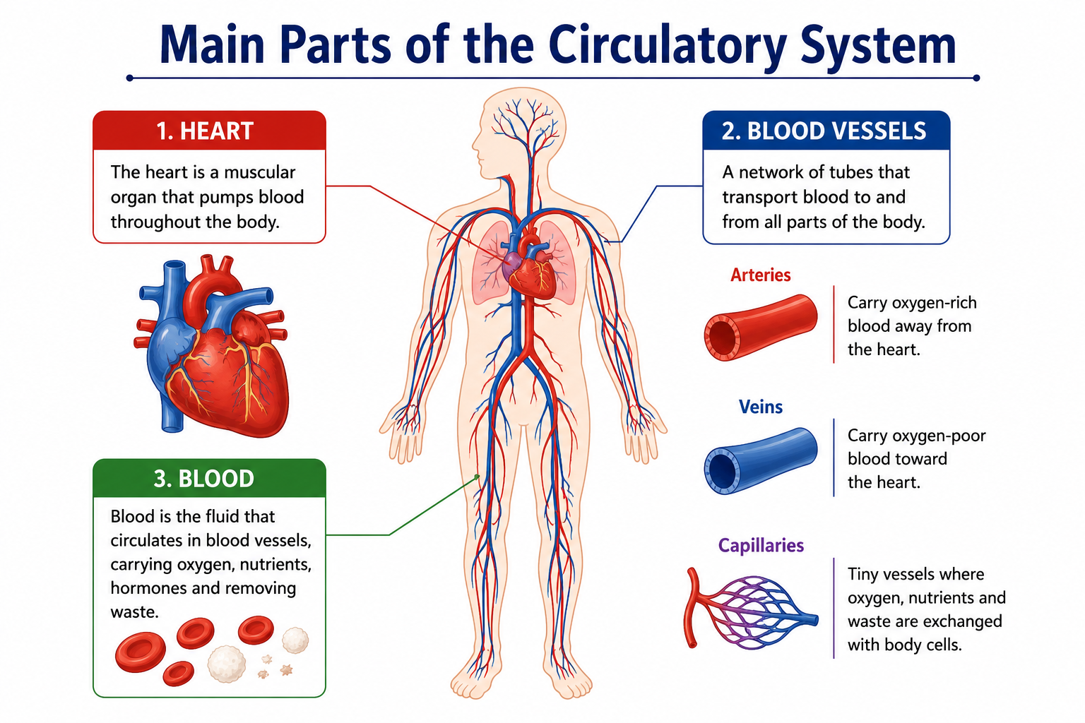
The circulatory system has three essential components that work as a unified team:
Heart
The heart is the muscular pump at the center of everything. Located slightly left of center in your chest, behind the sternum (breastbone), this fist-sized organ generates the force needed to push blood through approximately 60,000 miles (96,000 km) of blood vessels in the human body. Yes, if you stretched all your blood vessels end to end, they’d circle Earth more than twice.
Blood
Blood is the actual transport medium—a liquid tissue that carries oxygen, nutrients, hormones, waste products, and immune cells throughout the body. An average adult has about 4.7 to 5.5 liters of blood, which makes up roughly 7–8% of total body weight.
Blood Vessels
These are the tubes through which blood travels. There are three main types—arteries, veins, and capillaries—each designed with a specific structure suited to its function.
Structure of the Human Heart
The heart is a hollow, muscular organ. Its outer wall is called the myocardium (heart muscle), and it’s surrounded by a protective double-layered sac called the pericardium. The inner lining is called the endocardium.
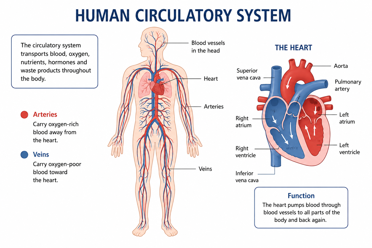
🫀 HEART ANATOMY DIAGRAM (Descriptive)

                  [Superior Vena Cava]
                         |
         [Right Atrium]──────[Left Atrium]
              |                    |
         [Tricuspid             [Mitral
          Valve]                 Valve]
              |                    |
         [Right Ventricle]──[Left Ventricle]
              |                    |
    [Pulmonary Artery]        [Aorta]
              |                    |
           [Lungs]              [Body]
Chambers of the Heart
The heart is divided into four chambers:
| Chamber | Location | Function |
|---|---|---|
| Right Atrium | Upper right | Receives deoxygenated blood from the body via vena cava |
| Right Ventricle | Lower right | Pumps deoxygenated blood to the lungs via pulmonary artery |
| Left Atrium | Upper left | Receives oxygenated blood from the lungs via pulmonary veins |
| Left Ventricle | Lower left | Pumps oxygenated blood to the entire body via the aorta |
Quick Tip: The left ventricle has the thickest wall of all four chambers because it has to push blood all the way to your toes and fingertips—much more work than pumping blood just to the nearby lungs.
A wall called the septum divides the heart into left and right halves, ensuring oxygenated and deoxygenated blood never mix in a healthy heart.
Heart Valves
Valves act like one-way doors inside the heart, preventing blood from flowing backward. There are four main valves:
- Tricuspid Valve — between right atrium and right ventricle (3 leaflets)
- Pulmonary Valve — between right ventricle and pulmonary artery
- Mitral (Bicuspid) Valve — between left atrium and left ventricle (2 leaflets)
- Aortic Valve — between left ventricle and aorta
The “lub-dub” sound of a heartbeat is actually the sound of these valves snapping shut. “Lub” is the closing of the tricuspid and mitral valves; “dub” is the closing of the pulmonary and aortic valves.
Major Blood Vessels Connected to the Heart
- Aorta — the largest artery; carries oxygenated blood from the left ventricle to the body
- Pulmonary Artery — carries deoxygenated blood from the right ventricle to the lungs
- Pulmonary Veins — carry oxygenated blood from the lungs back to the left atrium (there are four of them)
- Superior Vena Cava — brings deoxygenated blood from the upper body to the right atrium
- Inferior Vena Cava — brings deoxygenated blood from the lower body to the right atrium
Coronary Arteries
The heart muscle itself needs its own blood supply—it doesn’t absorb oxygen directly from the blood flowing through its chambers. The coronary arteries branch off the aorta and wrap around the heart to supply it with oxygen-rich blood. There are two main coronary arteries: the left coronary artery and the right coronary artery. A blockage in these arteries leads to a heart attack.
Types of Blood Vessels
Arteries
Arteries carry blood away from the heart (remember: Arteries = Away). With only a few exceptions, arteries carry oxygenated blood. They have thick, elastic walls to withstand the high pressure generated by the pumping heart. The largest artery in the body is the aorta.
Arteries branch into smaller vessels called arterioles, which then connect to capillaries.
Veins
Veins carry blood back toward the heart. They generally carry deoxygenated blood (again, with the exception of pulmonary veins). Their walls are thinner than arteries because blood pressure is much lower by the time it reaches them. Many veins, especially in the legs, contain valves to prevent blood from pooling backward due to gravity.
Veins begin as small venules that collect blood from capillaries and merge into larger veins.
Capillaries
Capillaries are the smallest blood vessels in the body—so narrow that red blood cells must travel through them single file. Their walls are just one cell thick, which makes them perfectly designed for exchange: oxygen and nutrients pass out into surrounding tissues, while carbon dioxide and waste products pass in. This exchange is the entire point of the circulatory system.
Arteries vs Veins vs Capillaries (Comparison Table)
| Feature | Arteries | Veins | Capillaries |
|---|---|---|---|
| Direction of Flow | Away from heart | Toward heart | Between arteries & veins |
| Blood Type (usually) | Oxygenated | Deoxygenated | Mixed |
| Wall Thickness | Thick, muscular | Thin, less muscular | Single cell layer |
| Pressure | High | Low | Very low |
| Valves | No (except semilunar) | Yes (in limbs) | No |
| Lumen Size | Narrow | Wide | Extremely narrow |
| Pulse | Present | Absent | Absent |
| Examples | Aorta, coronary artery | Vena cava, jugular vein | Tissue capillaries |
Types of Blood
Oxygenated Blood
Oxygenated blood is blood that is rich in oxygen. It flows from the lungs to the left side of the heart and then out to the rest of the body through the arteries. It appears bright red because of the oxygen-bound hemoglobin in red blood cells.
Deoxygenated Blood
Deoxygenated blood has released most of its oxygen to body tissues and is now carrying carbon dioxide waste back to the lungs. It flows through the veins to the right side of the heart and then to the lungs. It appears darker red (often described as purplish-red) rather than the vivid red of oxygenated blood.
Common Misconception: Many students think deoxygenated blood is actually blue. It’s not. It’s dark red. Blood only appears blue through your skin because of how light penetrates and reflects through tissue. In textbook diagrams, blue is just a color convention used to distinguish deoxygenated blood from oxygenated blood.
Components of Blood
Blood is far more complex than it looks. Let’s break down what it’s actually made of:
Red Blood Cells (RBCs)
Also called erythrocytes, RBCs are the most numerous blood cells. Their main job is to transport oxygen from the lungs to tissues and carry some carbon dioxide back. They contain hemoglobin, an iron-containing protein that binds oxygen. Mature human RBCs have no nucleus, which gives them more room to carry hemoglobin. They live for about 120 days.
Normal RBC count:
- Men: 4.7–6.1 million cells/µL
- Women: 4.2–5.4 million cells/µL
White Blood Cells (WBCs)
Also called leukocytes, WBCs are the soldiers of the immune system. They come in several types—neutrophils, lymphocytes, monocytes, eosinophils, and basophils—each with a specific role in fighting bacteria, viruses, parasites, and even cancer cells. Unlike RBCs, WBCs do have nuclei.
Normal WBC count: 4,500–11,000 cells/µL
Platelets
Platelets (thrombocytes) are tiny cell fragments that play a critical role in blood clotting. When you cut yourself, platelets rush to the site, clump together, and form a plug to stop bleeding. This process, combined with clotting proteins in plasma, creates a blood clot.
Normal platelet count: 150,000–400,000/µL
Plasma
Plasma is the liquid portion of blood—a pale yellowish fluid that makes up about 55% of total blood volume. It’s mostly water (about 90%) but also contains dissolved proteins (like albumin, fibrinogen, and immunoglobulins), electrolytes, hormones, nutrients, and waste products.
Blood Components Summary Table
| Component | Percentage of Blood | Main Function |
|---|---|---|
| Plasma | ~55% | Transport medium; carries nutrients, hormones, waste |
| Red Blood Cells | ~44% | Oxygen transport |
| White Blood Cells | <1% | Immune defense |
| Platelets | <1% | Blood clotting |
Blood Groups and Rh Factor
Human blood is classified into four main ABO blood groups based on the antigens present on the surface of red blood cells:
| Blood Group | Antigen on RBC | Antibody in Plasma | Can Donate To | Can Receive From |
|---|---|---|---|---|
| A | A antigen | Anti-B | A, AB | A, O |
| B | B antigen | Anti-A | B, AB | B, O |
| AB | A and B antigens | None | AB only | A, B, AB, O |
| O | No antigen | Anti-A and Anti-B | A, B, AB, O | O only |
The Rh factor is another antigen found on RBCs. If you have it, you’re Rh positive (+); if you don’t, you’re Rh negative (−). This is why blood types are written as A+, O−, AB+, etc.
Universal Donor: O negative (O−)
Universal Recipient: AB positive (AB+)
Double Circulation Explained
Humans (and other mammals) have what’s called a double circulation system. This means blood passes through the heart twice for every complete circuit of the body. The heart is divided into two separate pumping systems that work simultaneously.
📊 CIRCULATION FLOW DIAGRAM

PULMONARY CIRCULATION:
Right Ventricle → Pulmonary Artery → Lungs → Pulmonary Veins → Left Atrium

SYSTEMIC CIRCULATION:
Left Ventricle → Aorta → Body Tissues → Vena Cava → Right Atrium
Pulmonary Circulation
The pulmonary circuit is the shorter loop. It carries deoxygenated blood from the right ventricle to the lungs, where carbon dioxide is released and oxygen is absorbed. The freshly oxygenated blood then returns to the left atrium via the pulmonary veins. This entire loop covers a short distance—just heart to lungs and back.
Systemic Circulation
The systemic circuit is the longer, more extensive loop. It carries oxygenated blood from the left ventricle out through the aorta to every organ and tissue in the body. After delivering oxygen and nutrients, the blood collects in veins and returns to the right atrium via the superior and inferior vena cava.
Why Double Circulation? A single-circuit system (like fish have) means oxygenated and deoxygenated blood mix, reducing efficiency. Double circulation keeps them separate, ensuring organs always receive fully oxygenated blood at adequate pressure.
Blood Flow Through the Heart (Step-by-Step)
Here’s the complete journey of blood through your heart, step by step:
- Deoxygenated blood from the upper body enters the right atrium via the superior vena cava.
- Deoxygenated blood from the lower body enters the right atrium via the inferior vena cava.
- The right atrium contracts, pushing blood through the tricuspid valve into the right ventricle.
- The right ventricle contracts, pushing blood through the pulmonary valve into the pulmonary artery.
- Blood travels to the lungs, where it releases CO₂ and picks up oxygen (gas exchange).
- Oxygenated blood returns from the lungs via four pulmonary veins into the left atrium.
- The left atrium contracts, pushing blood through the mitral (bicuspid) valve into the left ventricle.
- The left ventricle contracts powerfully, pushing blood through the aortic valve into the aorta.
- Blood travels through the aorta and its branches to all parts of the body.
- After delivering oxygen, blood collects in veins and returns to the right atrium—and the cycle repeats.
Cardiac Cycle Explained
The cardiac cycle refers to the sequence of events that occurs with each heartbeat. It includes two main phases:
Systole
Systole is the phase of contraction. When the ventricles contract (ventricular systole), blood is forcefully ejected into the pulmonary artery and aorta. During atrial systole, the atria contract to fill the ventricles. The blood pressure measured during systole is the top number in a blood pressure reading.
Diastole
Diastole is the phase of relaxation. The heart muscle relaxes, and the chambers refill with blood. The valves prevent blood from flowing back. The blood pressure during diastole is the bottom number in a blood pressure reading.
One complete cardiac cycle takes approximately 0.8 seconds at a resting heart rate of 75 beats per minute.
Heartbeat and Pulse
The heartbeat is initiated by a specialized group of cells called the sinoatrial (SA) node, located in the wall of the right atrium. It’s often called the natural pacemaker of the heart because it generates the electrical signal that triggers each heartbeat.
The signal travels through:
SA Node → AV Node (atrioventricular node) → Bundle of His → Purkinje Fibers → Ventricles contract
This electrical conduction system ensures the heart beats in a coordinated, rhythmic pattern.
Normal resting heart rate: 60–100 beats per minute (bpm)
- Bradycardia: < 60 bpm
- Tachycardia: > 100 bpm
Pulse is the rhythmic expansion of an artery felt at certain points on the body (wrist, neck, temple) corresponding to each heartbeat. It’s a quick way to assess heart rate.
Blood Pressure Explained
Blood pressure is the force that blood exerts against the walls of blood vessels. It’s measured in millimeters of mercury (mmHg) and written as two numbers: systolic/diastolic.
Normal Blood Pressure
Normal: Less than 120/80 mmHg
High Blood Pressure (Hypertension)
- Elevated: 120–129/less than 80
- Stage 1 Hypertension: 130–139/80–89
- Stage 2 Hypertension: 140+/90+
Chronic high blood pressure damages blood vessel walls, strains the heart, and significantly increases the risk of heart attack and stroke.
Low Blood Pressure (Hypotension)
Defined as readings consistently below 90/60 mmHg. Symptoms include dizziness, fainting, and fatigue. While less commonly discussed, it can also be dangerous.
Functions of the Circulatory System
The circulatory system does much more than just move blood around. Here’s a comprehensive list:
Oxygen delivery — Transports O₂ from lungs to all body cells
Carbon dioxide removal — Carries CO₂ from cells back to the lungs
Nutrient transport — Delivers glucose, amino acids, fatty acids from the digestive system
Waste removal — Carries metabolic waste to kidneys and liver for elimination
Hormone transport — Carries hormones from glands to target organs
Immune defense — Transports WBCs and antibodies throughout the body
Temperature regulation — Redirects blood flow to manage body temperature
pH regulation — Helps maintain the acid-base balance of body fluids
Fluid balance — Regulates fluid distribution between tissues and blood
Diseases of the Circulatory System
Understanding what can go wrong helps you appreciate how everything works when it goes right.
Hypertension
Persistently elevated blood pressure that forces the heart to work harder. It’s often called the “silent killer” because it has no obvious symptoms until serious damage has occurred. Risk factors include a high-salt diet, obesity, stress, smoking, and genetic predisposition.
Atherosclerosis
This is the buildup of fatty deposits called plaques inside artery walls. Over time, plaques harden and narrow the arteries, reducing blood flow. It’s the underlying cause of most heart attacks and many strokes.
Heart Attack (Myocardial Infarction)
A heart attack occurs when blood flow to a section of the heart muscle is completely blocked—usually by a blood clot forming on top of a ruptured atherosclerotic plaque. Without blood, that section of heart muscle begins to die. Symptoms include chest pain, shortness of breath, and pain radiating to the left arm.
Stroke
A stroke happens when blood flow to part of the brain is cut off—either by a blocked artery (ischemic stroke, the most common type) or a burst blood vessel (hemorrhagic stroke). Brain cells die rapidly, causing lasting neurological damage.
Anemia
Anemia occurs when the blood doesn’t have enough healthy red blood cells or hemoglobin to carry adequate oxygen. Symptoms include fatigue, pale skin, and weakness. It can result from iron deficiency, vitamin B12 deficiency, blood loss, or underlying disease.
How to Keep Your Heart Healthy
Your lifestyle choices have a massive impact on cardiovascular health. Here’s what the science consistently supports:
- Exercise regularly — Aim for at least 150 minutes of moderate aerobic activity per week
- Eat a heart-healthy diet — Emphasize fruits, vegetables, whole grains, and healthy fats (Mediterranean diet is well-supported)
- Don’t smoke — Smoking damages blood vessel walls and accelerates atherosclerosis
- Manage stress — Chronic stress raises blood pressure and promotes inflammation
- Get enough sleep — Poor sleep is linked to hypertension and increased heart disease risk
- Manage existing conditions — Control diabetes, high cholesterol, and high blood pressure with medical help
- Maintain a healthy weight — Excess body fat, especially around the abdomen, strains the heart
- Get regular check-ups — Know your blood pressure, cholesterol, and blood sugar numbers
Common Circulatory System Terms Every Student Should Know
| Term | Definition |
|---|---|
| Cardiac Output | Volume of blood pumped by the heart per minute (heart rate × stroke volume) |
| Stroke Volume | Amount of blood pumped per heartbeat |
| Hemoglobin | Iron-containing protein in RBCs that carries oxygen |
| Erythropoiesis | Production of red blood cells (occurs in bone marrow) |
| Hematocrit | Percentage of blood volume occupied by RBCs |
| Fibrinogen | A plasma protein involved in blood clotting |
| Vasoconstriction | Narrowing of blood vessels |
| Vasodilation | Widening of blood vessels |
| Tachycardia | Heart rate above 100 bpm |
| Bradycardia | Heart rate below 60 bpm |
| Ischemia | Insufficient blood supply to a tissue |
| Pericardium | The protective sac surrounding the heart |
| Endocardium | The inner lining of the heart chambers |
| Myocardium | The heart muscle itself |
| Sphygmomanometer | Instrument used to measure blood pressure |
Common Mistakes Students Make
Even sharp students fall into predictable traps when studying the circulatory system. Here are the most common ones—and how to avoid them:
- Confusing arteries and veins by their oxygen content. Arteries carry blood away from the heart; veins carry blood toward the heart. The pulmonary artery carries deoxygenated blood, and pulmonary veins carry oxygenated blood—this trips up nearly every student the first time.
- Thinking deoxygenated blood is blue. It’s not. It’s dark red. Blue is just a diagram convention.
- Mixing up systole and diastole. Systole = squeeze (contraction), Diastole = relax. Think of “systole starts with S for Squeeze.”
- Forgetting that the left ventricle has the thickest wall and pumping the most work.
- Confusing the SA node and AV node. The SA node starts the electrical signal; the AV node briefly delays it to allow the ventricles to fill before contracting.
- Ignoring pulmonary veins. Students often forget there are four pulmonary veins (two from each lung), not just one.
- Mixing up blood groups. Practice the donor/recipient table until it’s second nature.
Best Tips to Study the Circulatory System
🎯 Exam Tips Box
- Draw it, don’t just read it. Sketch the heart chambers and label them from memory. Repeat this daily for a week.
- Use color coding — red for oxygenated, blue for deoxygenated, even in your own notes.
- Create a flow chart of blood through the heart and body. Tracing a drop of blood from vena cava back to vena cava should become automatic.
- Make flashcards for valve names, vessel names, and heart sounds.
- Connect concepts — understand WHY each structure is shaped the way it is (thick left ventricle wall = more pumping demand).
- Test yourself using the practice questions at the end of this guide.
- Teach it — explain the cardiac cycle to a friend or even to yourself in a mirror. If you can teach it, you know it.
- Use mnemonics — “Try Pulling My Aorta” for valve order: Tricuspid, Pulmonary, Mitral, Aortic.
Human Circulatory System Practice Questions
20 Multiple Choice Questions (MCQs) with Answers
1. Which chamber of the heart pumps blood to the entire body?
- A) Right Atrium
- B) Left Atrium
- C) Right Ventricle
- D) Left Ventricle ✓
2. Which blood vessel carries oxygenated blood from the lungs to the heart?
- A) Pulmonary Artery
- B) Aorta
- C) Pulmonary Veins ✓
- D) Vena Cava
3. What is the normal resting heart rate for an adult?
- A) 40–60 bpm
- B) 60–100 bpm ✓
- C) 100–120 bpm
- D) 30–50 bpm
4. The “pacemaker” of the heart is the:
- A) SA Node ✓
- B) AV Node
- C) Bundle of His
- D) Purkinje Fibers
5. Which blood component is responsible for clotting?
- A) Red Blood Cells
- B) White Blood Cells
- C) Platelets ✓
- D) Plasma
6. The largest artery in the human body is the:
- A) Aorta ✓
- B) Pulmonary Artery
- C) Coronary Artery
- D) Femoral Artery
7. What percentage of blood volume does plasma make up?
- A) 25%
- B) 44%
- C) 55% ✓
- D) 70%
8. Which blood group is the universal donor?
- A) AB+
- B) A−
- C) O− ✓
- D) B+
9. The valve between the left atrium and left ventricle is the:
- A) Tricuspid Valve
- B) Aortic Valve
- C) Mitral (Bicuspid) Valve ✓
- D) Pulmonary Valve
10. Pulmonary circulation refers to blood flow between:
- A) Heart and Lungs ✓
- B) Heart and Brain
- C) Heart and Kidneys
- D) Lungs and Body
11. Which type of blood vessel has walls just one cell thick?
- A) Arteries
- B) Veins
- C) Capillaries ✓
- D) Arterioles
12. What does the term “diastole” refer to?
- A) Heart contraction
- B) Heart relaxation ✓
- C) Blood clot formation
- D) Valve closure
13. The iron-containing protein in red blood cells is called:
- A) Fibrinogen
- B) Hemoglobin ✓
- C) Albumin
- D) Immunoglobulin
14. Normal adult blood pressure is approximately:
- A) 140/90 mmHg
- B) 120/80 mmHg ✓
- C) 100/60 mmHg
- D) 160/100 mmHg
15. RBCs in humans live for approximately:
- A) 30 days
- B) 60 days
- C) 120 days ✓
- D) 200 days
16. Which blood cells are part of the immune system?
- A) Erythrocytes
- B) Thrombocytes
- C) Leukocytes ✓
- D) Platelets
17. Atherosclerosis is the buildup of:
- A) Calcium in bones
- B) Fatty plaques in arteries ✓
- C) Blood clots in veins
- D) Fluid in the pericardium
18. How much blood does an average adult have?
- A) 1–2 liters
- B) 3–4 liters
- C) 4.7–5.5 liters ✓
- D) 7–8 liters
19. The “lub” sound of the heartbeat is produced by the closure of:
- A) Aortic and Pulmonary valves
- B) Mitral and Tricuspid valves ✓
- C) Coronary valves
- D) Semilunar valves
20. What is the term for a heart rate below 60 bpm?
- A) Tachycardia
- B) Bradycardia ✓
- C) Arrhythmia
- D) Fibrillation
10 Short Answer Questions
- What is the difference between pulmonary and systemic circulation?
- Name the four chambers of the heart and their functions.
- What are the three types of blood vessels? How do their structures differ?
- What is the role of hemoglobin in red blood cells?
- Explain why the left ventricle has a thicker wall than the right ventricle.
- What is the cardiac cycle, and how long does one cycle take at rest?
- What is the difference between systole and diastole?
- What is atherosclerosis, and how does it lead to heart attacks?
- Define blood pressure and state the normal range for adults.
- What is the function of platelets in the blood?
5 Long Answer Questions
- Describe the complete path of a red blood cell from the right atrium through pulmonary circulation and back through systemic circulation to a cell in the left leg. Include all chambers, vessels, and valves encountered.
- Compare and contrast arteries, veins, and capillaries in terms of structure, function, direction of blood flow, pressure, and examples in the body.
- Explain the cardiac cycle in detail, including the roles of the SA node, AV node, Bundle of His, and Purkinje fibers in coordinating the heartbeat.
- Discuss the major components of blood—red blood cells, white blood cells, platelets, and plasma—and explain how each contributes to the overall functioning of the circulatory system.
- What are the main diseases of the circulatory system? For each disease you mention, explain its cause, mechanism, and potential prevention or treatment strategies.
Revision Checklist
Use this before your exam to make sure you haven’t missed anything important:
- I can name and describe the four chambers of the heart
- I can explain the function of all four heart valves
- I can trace blood flow through the entire circulatory system
- I understand the difference between pulmonary and systemic circulation
- I can explain double circulation and why it’s beneficial
- I know the structure and function of arteries, veins, and capillaries
- I can describe all four components of blood and their functions
- I understand the ABO blood group system and Rh factor
- I can explain the cardiac cycle, including systole and diastole
- I understand the role of the SA node as the heart’s pacemaker
- I know normal values for heart rate and blood pressure
- I can describe at least five diseases of the circulatory system
- I understand how lifestyle factors affect cardiovascular health
- I’ve completed all practice MCQs and checked my answers
- I can draw and label a basic diagram of the heart
Best Books for Learning Human Anatomy
Here are some excellent textbooks that biology teachers, nursing students, and medical exam candidates consistently recommend:
- “Human Anatomy & Physiology” by Marieb & Hoehn — Arguably the most widely used university-level A&P textbook. Clear, well-illustrated, and thorough.
- “Gray’s Anatomy for Students” by Drake, Vogl & Mitchell — The student edition of the legendary clinical anatomy reference. Excellent for detail-oriented learners.
- “Guyton and Hall Textbook of Medical Physiology” — The gold standard for understanding physiology, including cardiovascular function. Ideal for medical students.
- “The Anatomy Coloring Book” by Kapit & Elson — Don’t let the word “coloring” fool you—this is a serious learning tool that uses active engagement to reinforce anatomy.
- “Ross & Wilson Anatomy and Physiology in Health and Illness” — Popular among nursing students; clinical applications are highlighted throughout.
Free Online Anatomy Resources
You don’t need expensive textbooks to get started. These free, reputable resources are excellent:
- OpenStax Anatomy & Physiology — A completely free, peer-reviewed university-level textbook with excellent circulatory system chapters.
- Khan Academy Biology — Free video lessons and practice exercises on the circulatory system, heart anatomy, and blood.
- Biology LibreTexts — Open-access library of biology textbooks and articles, including detailed cardiovascular physiology.
- National Heart, Lung, and Blood Institute (NHLBI) — Reliable, medically reviewed information about how the heart works and common heart conditions.
- MedlinePlus — Maintained by the U.S. National Library of Medicine; excellent plain-language explanations of circulatory diseases.
Related Articles on LearnMinto
If you found this guide useful, these related articles will help you build a complete picture of human biology:
- 📖 Biology Study Guide — Your comprehensive overview of all major biology topics
- 📖 Human Anatomy Study Guide — A full tour of every major body system
- 📖 Cell Biology Study Guide — Understand the building blocks of life
- 📖 Human Respiratory System Study Guide — Learn how breathing and gas exchange work
- 📖 Human Digestive System Study Guide — Follow nutrients from your mouth to your cells
Frequently Asked Questions
Q1: What is the human circulatory system?
The human circulatory system is a network of the heart, blood vessels (arteries, veins, and capillaries), and blood that works together to deliver oxygen, nutrients, and hormones to body cells while removing carbon dioxide and waste products.
Q2: How many times does the heart beat in a day?
At an average resting rate of 70–75 beats per minute, the heart beats approximately 100,000 times per day and over 2.5 billion times in an average lifetime.
Q3: What is the difference between pulmonary and systemic circulation?
Pulmonary circulation carries deoxygenated blood from the right ventricle to the lungs and returns oxygenated blood to the left atrium. Systemic circulation carries oxygenated blood from the left ventricle to all body tissues and returns deoxygenated blood to the right atrium.
Q4: Why do arteries have thicker walls than veins?
Arteries receive blood directly from the heart under high pressure. Their thick, elastic, muscular walls allow them to withstand and manage this pressure. Veins carry blood at much lower pressure on its way back to the heart, so they don’t need walls as strong.
Q5: What is the universal blood donor type?
Blood type O negative (O−) is the universal donor because it has no A, B, or Rh antigens on red blood cells, meaning it won’t trigger an immune reaction in recipients of any blood type. This makes it critically important in emergency transfusions when there’s no time to test the patient’s blood type.
Q6: What causes a heart attack?
A heart attack (myocardial infarction) occurs when a coronary artery becomes completely blocked—usually by a blood clot that forms on a ruptured fatty plaque—cutting off oxygen supply to a section of heart muscle. Without oxygen, that muscle tissue begins to die within minutes.
Q7: What is blood pressure, and what is considered normal?
Blood pressure is the force blood exerts on artery walls. It’s recorded as two numbers: systolic pressure (during heartbeat) over diastolic pressure (between beats). A normal reading for adults is less than 120/80 mmHg.
Q8: How many liters of blood does the human body contain?
The average adult human body contains approximately 4.7 to 5.5 liters of blood. This can vary based on body size, sex, and health status.
Q9: What are the components of blood?
Blood is composed of four main components: red blood cells (carry oxygen), white blood cells (fight infection), platelets (aid clotting), and plasma (the liquid matrix that carries everything else).
Q10: What is the cardiac cycle?
The cardiac cycle refers to the complete sequence of events in one heartbeat—from the beginning of one contraction to the beginning of the next. It consists of systole (contraction phase) and diastole (relaxation phase) and lasts about 0.8 seconds at a resting heart rate.
Q11: What is the role of capillaries in the circulatory system?
Capillaries are the smallest and thinnest blood vessels, with walls just one cell thick. They’re the site of actual exchange between blood and body tissues—oxygen and nutrients diffuse out into cells, while carbon dioxide and waste diffuse into the blood.
Q12: Can the circulatory system repair itself after a heart attack?
The heart has very limited regenerative capacity. Cardiac muscle cells (cardiomyocytes) do not easily regenerate after death. Damaged heart tissue is typically replaced by scar tissue, which doesn’t contract. This is why preventing heart attacks through healthy lifestyle choices is so much more effective than treating them after the fact.
Summary
Let’s bring everything together. The human circulatory system is the body’s essential transport network, built around three core components: the heart, blood, and blood vessels.
The heart is a four-chambered muscular pump that drives two separate circuits simultaneously. The pulmonary circuit takes blood to the lungs for oxygen and removes CO₂; the systemic circuit delivers oxygenated blood to every tissue in the body. Blood passes through the heart twice per complete journey—which is why humans are said to have a double circulation.
Blood itself is a complex living tissue made of plasma, red blood cells, white blood cells, and platelets. Each component has essential roles in oxygen transport, immune defense, and injury response. Blood groups (ABO + Rh) are determined by antigens on RBC surfaces and matter enormously in transfusion medicine.
The cardiac cycle—driven by electrical signals from the SA node—coordinates the rhythmic contraction and relaxation of heart chambers. Each heartbeat produces measurable blood pressure, with 120/80 mmHg being the healthy benchmark for adults.
When this system is compromised—through hypertension, atherosclerosis, heart attacks, or anemia—the consequences affect every other body system. The good news is that lifestyle modifications like regular exercise, a healthy diet, not smoking, and managing stress are proven to protect cardiovascular health significantly.
Final Thoughts
The circulatory system is one of the most elegant and fascinating systems in the human body. Once you truly understand it—not just memorize the parts, but understand how and why each piece fits together—you’ll find that it becomes one of the easier topics to recall under exam pressure.
The key is building your knowledge layer by layer. Start with the big picture (what the system does), then zoom in to the anatomy (heart chambers, valves, vessels), then understand the processes (cardiac cycle, circulation routes), and finally connect it to real-life health (blood pressure, disease, prevention).
If you’re a student using this Human Circulatory System Study Guide for exam prep, work through the practice questions honestly—without peeking at answers first. If you’re a teacher, feel free to use the comparison tables, flowcharts, and revision checklist with your students.
Keep revisiting this guide as your course progresses. Biology builds on itself, and every time you come back, you’ll notice connections you didn’t see before. That’s the real sign that learning is happening.
Good luck, and keep your heart in it. Quite literally.
Disclaimer
This article is intended for educational and informational purposes only. While LearnMinto strives to provide accurate and up-to-date information, readers should verify important academic concepts through official textbooks, educational institutions, examination boards, or trusted scientific resources before relying on this content for exams or academic purposes. LearnMinto is not affiliated with any specific school, university, research institution, or examination board.