
Introduction
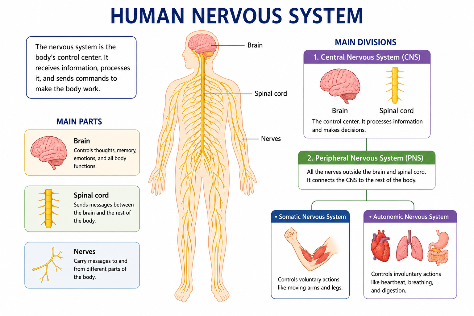
Have you ever yanked your hand away from a hot stove before you even consciously realized it was burning? Or felt your heart race before a big exam, your palms getting sweaty even though you’re just sitting at a desk? Both of those experiences are your nervous system working at full speed—processing information, triggering responses, and keeping you safe and functioning, all without asking your permission first.
The nervous system is arguably the most complex and fascinating system in the entire human body. It controls everything: your thoughts, memories, emotions, movements, reflexes, and your ability to sense the world around you. And yet, at its most fundamental level, it all comes down to billions of specialized cells passing electrical and chemical signals to one another at astonishing speed.
This Human Nervous System Study Guide is built for students who want a thorough, exam-ready understanding of how the nervous system works—from the big-picture structure of the brain down to the molecular events happening at a single synapse. Whether you’re a high school student tackling biology for the first time, a nursing student preparing for clinical concepts, or a medical entrance exam candidate looking to sharpen your knowledge, this guide covers everything you need.
We’ll explore the structure of the brain and spinal cord, understand how neurons generate and transmit signals, break down the reflex arc step by step, and look at what happens when this system breaks down due to disease. Along the way, you’ll find comparison tables, real-life examples, exam tips, and a full set of practice questions to test yourself before the big day.
Let’s get into it—your brain is literally built for this.
Key Takeaways
Before You Dive In — Key Takeaways
- The nervous system has two main divisions: the Central Nervous System (CNS) and the Peripheral Nervous System (PNS).
- The CNS consists of the brain and spinal cord; the PNS connects the CNS to the rest of the body.
- Neurons are the basic functional units of the nervous system; they come in three types: sensory, motor, and interneurons.
- Nerve impulses travel as electrical signals (action potentials) along neurons and as chemical signals across synapses.
- The brain has four main regions: the cerebrum, cerebellum, brainstem, and diencephalon (including thalamus and hypothalamus).
- A reflex action is a rapid, automatic response to a stimulus that does not require conscious thought.
- The autonomic nervous system controls involuntary functions and is divided into sympathetic (fight or flight) and parasympathetic (rest and digest) divisions.
- Major nervous system disorders include Alzheimer’s disease, Parkinson’s disease, multiple sclerosis, epilepsy, and stroke.
What Is the Human Nervous System?
The human nervous system is the body’s master communication and control network. It collects information from both inside and outside the body, processes that information, and coordinates appropriate responses—all within fractions of a second. It’s what allows you to read these words, feel the chair beneath you, notice a sound outside the window, and decide what to have for lunch, all more or less simultaneously.
Structurally, the nervous system is an incredibly dense web of nerve cells (neurons) and supporting cells (glial cells), connected into pathways, circuits, and networks. These networks extend from your brain down through your spinal cord and branch outward into every region of your body—including your skin, muscles, organs, and sensory receptors.
The nervous system works in close partnership with the endocrine system (which uses hormones for slower, longer-lasting communication), but where the endocrine system is like sending a letter, the nervous system is like making a phone call. Fast, direct, and specific.
💡 Important Fact: The human brain contains approximately 86 billion neurons, and each neuron can form thousands of connections with other neurons. The total number of synaptic connections in the brain is estimated to be around 100 trillion—more than the number of stars in the Milky Way galaxy.
Why Is the Nervous System Important?
It’s worth pausing to appreciate just how central the nervous system is to everything you do and experience:
- Without it, your lungs wouldn’t know to breathe, your heart wouldn’t maintain its rhythm, and your digestive system wouldn’t coordinate its movements.
- Your ability to sense pain is a nervous system function—one that’s lifesaving, because pain signals damage and drives you to protect injured areas.
- Every memory you’ve ever formed, every skill you’ve learned, every emotion you’ve felt—all of it exists as patterns of neural connections in your brain.
- It integrates sensory input from your eyes, ears, nose, skin, and internal organs, allowing you to build a real-time model of your environment and respond to it intelligently.
From an academic perspective, the nervous system is one of the most heavily tested topics in biology, anatomy, physiology, and medical entrance examinations. It connects to nearly every other body system, which means understanding it well gives you a powerful foundation for the rest of your studies.
Main Parts of the Nervous System
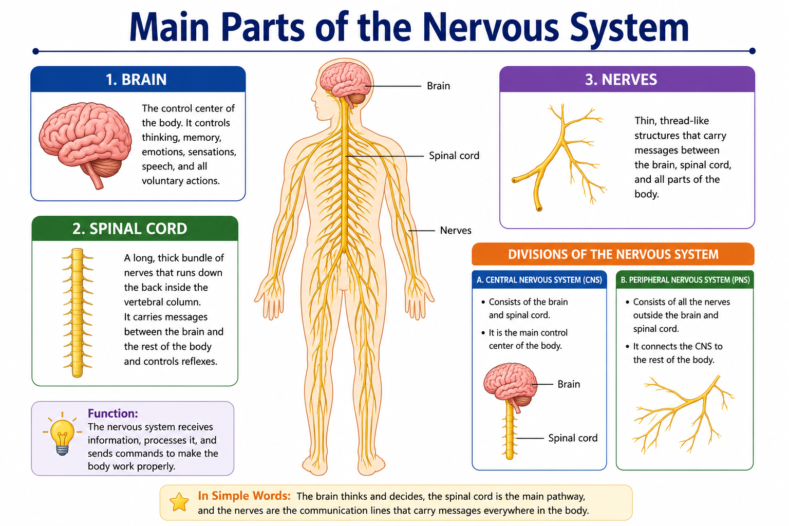
The nervous system is organized into two major divisions, with several important subdivisions beneath them
Central Nervous System (CNS)
The CNS is the processing hub of the entire nervous system. It consists of the brain and the spinal cord. All the information arriving from the body gets sent to the CNS, which analyzes it and sends out appropriate commands. The CNS is protected by bone (the skull protects the brain; the vertebral column protects the spinal cord) and by three layers of protective membranes called meninges, as well as a specialized fluid called cerebrospinal fluid (CSF) that cushions and nourishes nervous tissue.
Peripheral Nervous System (PNS)
The PNS is everything outside the CNS—the vast network of nerves that extends from the brain and spinal cord out to every part of the body. It’s the communication highway between the CNS and the muscles, glands, and sensory organs. Without the PNS, the brain and spinal cord would be isolated, completely unable to receive information or deliver commands.
Somatic Nervous System
The somatic nervous system handles voluntary activities—the movements you consciously control, like picking up a pen, speaking, or walking. It also carries sensory information (touch, temperature, pain, pressure) from the skin and muscles back to the CNS. It involves two types of nerve fibers: afferent fibers (carrying sensory signals toward the CNS) and efferent fibers (carrying motor signals away from the CNS to muscles).
Autonomic Nervous System
The autonomic nervous system (ANS) controls involuntary body functions—things that happen automatically without any conscious input from you. This includes your heartbeat, breathing rate, digestion, blood pressure, and glandular secretions. The ANS has two major branches that generally oppose each other to maintain balance.
Sympathetic Nervous System
Often described as the “fight or flight” system, the sympathetic division activates when you’re stressed, excited, or in danger. It speeds up your heart rate, dilates your airways, redirects blood to muscles, dilates your pupils, and slows digestion—essentially preparing your body for rapid physical action. The primary neurotransmitters involved are epinephrine (adrenaline) and norepinephrine (noradrenaline).
Parasympathetic Nervous System
The “rest and digest” system, the parasympathetic division, is active when you’re calm and relaxed. It slows the heart rate, promotes digestion, stimulates salivation, and generally conserves energy. Its primary neurotransmitter is acetylcholine. Together, the sympathetic and parasympathetic systems act like the accelerator and brake pedals of the body.
Structure of the Human Brain
The brain is the most complex organ in the known universe. Weighing about 1.4 kg (roughly 3 pounds) in an adult, it’s largely made of fatty tissue and contains an extraordinary density of neurons and their connections. Let’s look at the major structural regions:
🧠 BRAIN ANATOMY DIAGRAM (Descriptive)

              [Cerebrum - Large, wrinkled, upper portion]
              [Divides into Left and Right Hemispheres]
                        |
            [Four Lobes of the Cerebrum]
    [Frontal] [Parietal] [Temporal] [Occipital]
                        |
              [Thalamus & Hypothalamus]
              [Deep inside, above brainstem]
                        |
            [Cerebellum - Back, lower portion]
            [Controls balance and coordination]
                        |
               [Brainstem - Connects to Spinal Cord]
         [Midbrain] → [Pons] → [Medulla Oblongata]
Cerebrum
The cerebrum is the largest part of the brain, making up about 85% of total brain weight. Its outer layer, the cerebral cortex, is deeply folded into ridges (gyri) and grooves (sulci), which dramatically increases its surface area—allowing far more neurons to be packed in than if the surface were smooth.
The cerebrum is divided into two hemispheres—left and right—connected by a thick band of nerve fibers called the corpus callosum. Each hemisphere controls the opposite side of the body (the left hemisphere controls the right side, and vice versa).
The cerebrum is further divided into four lobes, each with distinct primary functions:
| Lobe | Location | Primary Functions |
|---|---|---|
| Frontal Lobe | Front of the brain | Thinking, planning, decision-making, voluntary movement, personality, speech production (Broca’s area) |
| Parietal Lobe | Top, behind frontal lobe | Processing touch, pressure, pain, temperature; spatial awareness |
| Temporal Lobe | Sides, behind temples | Hearing, language comprehension (Wernicke’s area), memory, emotion |
| Occipital Lobe | Back of the brain | Visual processing; interpreting what you see |
Cerebellum
Located at the back and bottom of the brain, the cerebellum is sometimes called the “little brain”—which is literally what its Latin name means. It coordinates muscle movements, maintains balance and posture, and refines motor skills. Without the cerebellum, movements would be jerky and uncoordinated. Athletes and musicians have particularly well-developed cerebellums due to years of precise movement practice.
Brainstem
The brainstem connects the brain to the spinal cord and is divided into three regions:
- Midbrain — involved in visual and auditory reflexes, eye movement
- Pons — connects the cerebellum to the brainstem; involved in sleep, breathing, and facial movement
- Medulla Oblongata — controls vital autonomic functions: heart rate, breathing, blood pressure, swallowing, and vomiting. It’s absolutely essential for survival.
Thalamus
Think of the thalamus as the brain’s relay station. Almost all sensory information (except smell) is routed through the thalamus before being sent to the appropriate area of the cerebral cortex for conscious processing. It acts as a filter and gatekeeper, deciding what sensory data deserves your attention.
Hypothalamus
The hypothalamus sits just below the thalamus and serves as the critical link between the nervous system and the endocrine (hormonal) system. It regulates body temperature, hunger, thirst, sleep cycles, and emotional responses. It controls the pituitary gland—the “master gland” of the body—making the hypothalamus one of the most functionally powerful small structures in the entire brain.
Structure of the Spinal Cord
The spinal cord is a long, cylindrical bundle of nerve tissue running from the brainstem down through the vertebral column, ending around the first or second lumbar vertebra. In adults, it’s approximately 45 cm long and about the thickness of a finger.
If you look at a cross-section of the spinal cord, you’ll notice two distinct regions:
- Gray Matter — shaped like a butterfly or the letter “H” in the center; contains the cell bodies of neurons
- White Matter — surrounds the gray matter; consists of myelinated axons that form ascending (sensory) and descending (motor) tracts
The spinal cord serves two major functions: it’s the pathway for information traveling up to the brain (sensory signals) and commands traveling down from the brain (motor signals). It also coordinates spinal reflexes independently, without needing input from the brain—which is exactly why you pull your hand away from something painful before you consciously register the pain.
What Are Neurons?
Neurons are the fundamental building blocks of the nervous system—the cells that actually carry the electrical and chemical signals that make all nervous system functions possible. The human body contains somewhere in the range of 86–100 billion neurons, though they come in several types and many shapes.
Structure of a Neuron
🔬 NEURON STRUCTURE DIAGRAM (Descriptive)

[Dendrites] → [Cell Body / Soma] → [Axon Hillock] → [Axon] → [Myelin Sheath]
                                                                      ↓
                                                           [Nodes of Ranvier]
                                                                      ↓
                                                         [Axon Terminal / Synaptic Knob]
                                                                      ↓
                                              [Synapse → Next Neuron or Effector]
A typical neuron has three main structural components:
- Cell Body (Soma) — contains the nucleus and organelles; the metabolic center of the neuron
- Dendrites — short, branching extensions that receive signals from other neurons and carry them toward the cell body
- Axon — a single, long projection that carries the electrical signal away from the cell body toward another neuron, muscle, or gland
Many axons are wrapped in a fatty insulating layer called the myelin sheath, produced by specialized cells called Schwann cells (in the PNS) or oligodendrocytes (in the CNS). The myelin sheath dramatically speeds up signal transmission. The gaps in the myelin sheath are called Nodes of Ranvier, and the signal effectively “jumps” between these nodes in a process called saltatory conduction.
At the end of the axon are axon terminals (synaptic knobs), which release chemical messengers called neurotransmitters into the gap between neurons (the synapse).
Types of Neurons
Sensory Neurons
Also called afferent neurons, sensory neurons carry information from sensory receptors (in the skin, eyes, ears, nose, and internal organs) toward the CNS. They convert physical stimuli—heat, pressure, light, sound, chemicals—into electrical signals. When you touch something hot, it’s a sensory neuron that first picks up that information.
Motor Neurons
Also called efferent neurons, motor neurons carry commands from the CNS out to muscles and glands (the effectors). When your brain decides to move your arm, it’s motor neurons that deliver that instruction to the appropriate muscles. They are the “output” neurons of the nervous system.
Interneurons
Interneurons (also called relay neurons or association neurons) are found exclusively within the CNS. They connect sensory and motor neurons, process information, and form the complex networks responsible for thought, memory, learning, and decision-making. The vast majority of neurons in the brain are interneurons.
Parts of a Neuron and Their Functions (Comparison Table)
| Structure | Location | Function |
|---|---|---|
| Dendrites | Extending from cell body | Receive incoming signals from other neurons |
| Cell Body (Soma) | Central part of neuron | Houses nucleus; integrates incoming signals |
| Axon Hillock | Where axon meets cell body | Decides whether to fire an action potential |
| Axon | Extends from cell body | Conducts electrical signal away from cell body |
| Myelin Sheath | Wraps around axon | Insulates axon; speeds up signal transmission |
| Nodes of Ranvier | Gaps in myelin sheath | Allows saltatory (jumping) conduction |
| Axon Terminal | End of axon | Releases neurotransmitters into the synapse |
| Synapse | Gap between neurons | Chemical transmission point between neurons |
How Nerve Impulses Travel
Understanding how signals actually move through neurons is one of the trickier parts of nervous system biology—but once it clicks, it’s genuinely fascinating. The whole process involves electricity and chemistry working together.
Resting Potential
When a neuron is not actively sending a signal, it’s in a resting state. At rest, the inside of the neuron is negatively charged relative to the outside (approximately −70 mV). This charge difference is maintained by the unequal distribution of ions—particularly sodium (Na⁺) and potassium (K⁺)—across the cell membrane, maintained by the sodium-potassium pump. This state of readiness is called the resting membrane potential.
Action Potential
When a neuron is sufficiently stimulated, the membrane’s permeability changes rapidly:
- Depolarization — Sodium channels open; Na⁺ ions rush into the cell, making the inside more positive (the membrane potential shoots up to about +30 mV)
- Repolarization — Sodium channels close; potassium channels open; K⁺ ions rush out, restoring the negative charge inside
- Refractory Period — A brief period during which the neuron cannot fire again; ensures signals travel in only one direction
This rapid electrical change—the action potential—travels along the axon like a wave, from the cell body toward the axon terminal. In myelinated neurons, this process is faster because the action potential jumps from node to node (saltatory conduction), reaching speeds of up to 120 meters per second.
Synaptic Transmission
When the action potential reaches the axon terminal, it triggers the release of neurotransmitters from tiny membrane packages called synaptic vesicles. These neurotransmitters cross the narrow synaptic cleft (the gap between two neurons) and bind to receptor proteins on the next neuron’s dendrites or cell body.
Depending on the neurotransmitter and receptor type, this can either:
- Excite the next neuron (making it more likely to fire) — Excitatory Post-Synaptic Potential (EPSP)
- Inhibit the next neuron (making it less likely to fire) — Inhibitory Post-Synaptic Potential (IPSP)
Common neurotransmitters include:
- Acetylcholine — muscle activation, memory, attention
- Dopamine — reward, movement, motivation
- Serotonin — mood regulation, sleep, appetite
- GABA — the main inhibitory neurotransmitter; calms neural activity
- Glutamate — the main excitatory neurotransmitter; involved in learning and memory
- Norepinephrine — alertness, stress response
After binding, neurotransmitters are either broken down by enzymes, reabsorbed by the sending neuron (reuptake), or diffuse away—ending the signal.
Reflex Action Explained
A reflex is a rapid, automatic, and stereotyped response to a specific stimulus. The key word is automatic—reflexes don’t require conscious thought or decision-making. They’re hardwired responses designed for speed, because in many situations (like touching something dangerously hot), even a fraction of a second’s delay can mean serious injury.
Reflex Arc
The reflex arc is the neural pathway through which a reflex operates. It bypasses the brain for speed, routing through the spinal cord instead.
🔄 REFLEX ARC DIAGRAM (Descriptive)

STIMULUS (e.g., hot surface)
        ↓
SENSORY RECEPTOR (detects stimulus in skin)
        ↓
SENSORY (AFFERENT) NEURON (carries signal toward CNS)
        ↓
INTERNEURON in Spinal Cord (processes signal; no brain involvement for spinal reflexes)
        ↓
MOTOR (EFFERENT) NEURON (carries command away from CNS)
        ↓
EFFECTOR (muscle or gland responds)
        ↓
RESPONSE (e.g., hand withdraws)
The signal also travels up to the brain simultaneously—which is why you feel the pain and become aware of it a moment after your hand has already pulled away. The reflex happened independently.
Examples of Reflexes
- Withdrawal Reflex — pulling your hand away from a painful stimulus (like a pin or hot object)
- Knee-Jerk (Patellar) Reflex — tapping the tendon below the kneecap causes the lower leg to kick forward (a simple, two-neuron reflex with no interneuron)
- Pupillary Reflex — pupils constrict in bright light and dilate in dim light
- Gag Reflex — triggered when the back of the throat is touched
- Blinking Reflex — automatic closing of the eyelid in response to an object approaching the eye
Brain vs Spinal Cord (Comparison Table)
| Feature | Brain | Spinal Cord |
|---|---|---|
| Location | Inside the skull (cranium) | Inside the vertebral column |
| Protection | Skull + meninges + CSF | Vertebrae + meninges + CSF |
| Primary Role | Processing, thinking, coordination, consciousness | Relay station + spinal reflex center |
| Gray Matter Position | Outer layer (cortex) | Inner butterfly-shaped region |
| White Matter Position | Inner regions | Outer region surrounding gray matter |
| Weight/Size | ~1.4 kg; ~15 cm wide | ~45 cm long; ~1 cm wide |
| Controls | Voluntary and complex functions | Reflexes; relay of sensory/motor signals |
| Divisions | Cerebrum, cerebellum, brainstem, diencephalon | Cervical, thoracic, lumbar, sacral, coccygeal segments |
Types of Reflexes
Innate Reflexes
Innate reflexes (also called unconditioned reflexes) are reflexes you’re born with. They don’t require learning or experience—they’re genetically programmed and present from birth. Examples include the knee-jerk reflex, pupillary reflex, sucking reflex in newborns, and the withdrawal reflex. These reflexes are consistent across virtually all healthy individuals.
Conditioned Reflexes
First demonstrated by the Russian physiologist Ivan Pavlov in his famous experiments with dogs, conditioned reflexes (also called learned reflexes) are acquired through experience and repetition. Pavlov showed that dogs naturally salivated when food was presented (innate reflex), but after repeatedly pairing the food with the sound of a bell, the dogs eventually salivated at the bell’s sound alone—even without food being present. Conditioned reflexes form through association between a neutral stimulus and a biologically significant one.
Functions of the Nervous System
The nervous system performs an enormous variety of functions that can be organized into three broad categories:
- Sensory Input — Detecting changes (stimuli) inside and outside the body through sensory receptors; collecting data about the environment
- Integration — Processing and interpreting sensory information; deciding on an appropriate response; this is the primary function of the CNS
- Motor Output — Sending commands to muscles and glands; producing movement, secretion, or other responses
Beyond these three, the nervous system is specifically responsible for:
- Regulating all other organ systems (cardiovascular, respiratory, digestive, etc.)
- Enabling conscious thought, reasoning, and problem-solving
- Forming and retrieving memories
- Processing and experiencing emotions
- Maintaining homeostasis (internal balance) through feedback loops
- Coordinating precise voluntary movements
- Managing the sleep-wake cycle
- Processing language, reading, and communication
Nervous System Disorders
The complexity of the nervous system also makes it vulnerable. When neurons are damaged or die, the effects can be profound, because unlike many other body cells, most mature neurons cannot regenerate.
Alzheimer’s Disease
Alzheimer’s is a progressive neurodegenerative disease that slowly destroys memory and cognitive function. It’s characterized by the accumulation of amyloid plaques and tau protein tangles in the brain, particularly in the hippocampus (memory center) first, then spreading to other regions. It’s the most common cause of dementia worldwide and currently has no cure, though some medications can temporarily slow progression.
Parkinson’s Disease
Parkinson’s disease results from the progressive degeneration of neurons in the substantia nigra, a region of the midbrain that produces dopamine. Dopamine is essential for coordinating smooth, controlled movements. Without it, the characteristic symptoms emerge: resting tremor (shaking at rest), rigidity, slow movement (bradykinesia), and balance problems. Dopamine-replacement medications like levodopa help manage symptoms.
Multiple Sclerosis (MS)
Multiple sclerosis is an autoimmune disease in which the body’s immune system mistakenly attacks and destroys the myelin sheath surrounding neurons in the CNS. Without myelin, nerve signals slow down or fail to travel properly, causing symptoms that vary widely depending on which areas of the CNS are affected: vision problems, muscle weakness, numbness, balance issues, and fatigue. MS can follow a relapsing-remitting pattern or progress steadily.
Epilepsy
Epilepsy is a neurological condition characterized by recurrent, unprovoked seizures—episodes of abnormal, excessive, or synchronous neuronal activity in the brain. Seizures can range from brief lapses of attention to violent convulsions, depending on how much of the brain is involved. Many people with epilepsy can control their seizures with anticonvulsant medications; some cases are amenable to surgery.
Stroke
A stroke occurs when blood supply to a part of the brain is interrupted—either by a blocked blood vessel (ischemic stroke) or a ruptured blood vessel (hemorrhagic stroke). Without blood, brain cells begin dying rapidly. The effects depend on which region of the brain is deprived: a stroke in the frontal lobe might impair movement or speech; in the occipital lobe, it might impair vision. Recognizing a stroke quickly using the FAST acronym (Face drooping, Arm weakness, Speech difficulty, Time to call emergency services) saves lives.
How to Keep Your Brain Healthy
Your brain is remarkably responsive to your lifestyle choices throughout your entire life. Neuroscientists now know that the brain maintains significant neuroplasticity—the ability to reorganize itself, form new connections, and even generate new neurons in certain regions (like the hippocampus)—well into adulthood.
Here are evidence-based habits that genuinely support brain health:
Regular aerobic exercise — Increases blood flow to the brain, promotes neurogenesis in the hippocampus, and reduces the risk of cognitive decline
Prioritize quality sleep — The brain uses sleep to consolidate memories and clear metabolic waste products (including amyloid proteins); chronic sleep deprivation impairs cognitive function significantly
Eat a brain-friendly diet — The Mediterranean diet (rich in olive oil, fish, vegetables, nuts, and whole grains) is consistently linked to better cognitive aging
Stay mentally active — Learning new skills, reading, puzzles, and intellectually stimulating activities support neural connections
Maintain social connections — Social engagement is one of the strongest protective factors against cognitive decline and dementia
Avoid smoking and excessive alcohol — Both are neurotoxic and accelerate brain aging
Manage vascular risk factors — Controlling high blood pressure, diabetes, and high cholesterol protects the brain’s blood supply
Manage chronic stress — Prolonged cortisol elevation damages hippocampal neurons and impairs memory formation
Common Nervous System Terms Every Student Should Know
| Term | Definition |
|---|---|
| Neuron | A nerve cell; the basic functional unit of the nervous system |
| Neurotransmitter | A chemical messenger released across a synapse |
| Synapse | The junction between two neurons where chemical signaling occurs |
| Action Potential | The electrical signal that travels along a neuron |
| Resting Potential | The electrical charge of an inactive neuron (~−70 mV) |
| Myelin Sheath | Fatty insulating layer that speeds up nerve signal transmission |
| Reflex Arc | The neural pathway involved in a reflex action |
| Meninges | Three protective membranes surrounding the brain and spinal cord |
| Cerebrospinal Fluid (CSF) | Clear fluid that cushions and nourishes the CNS |
| Neuroplasticity | The brain’s ability to reorganize and form new neural connections |
| Afferent | Carrying signals toward the CNS (sensory) |
| Efferent | Carrying signals away from the CNS (motor) |
| Ganglion | A cluster of neuron cell bodies in the PNS |
| Gray Matter | Neural tissue containing cell bodies; involved in processing |
| White Matter | Neural tissue containing myelinated axons; involved in transmission |
| Cortex | The outer layer of the cerebrum (cerebral cortex) |
| Hippocampus | Brain region crucial for memory formation; located in temporal lobe |
| Saltatory Conduction | The jumping of action potentials between Nodes of Ranvier |
Common Mistakes Students Make
Even motivated students make predictable errors when studying neuroscience. Being aware of these pitfalls ahead of time can save you a lot of confusion:
- Confusing afferent and efferent. Afferent = arriving at CNS (sensory); efferent = exiting CNS (motor). A helpful trick: Afferent = Arriving; Efferent = Exiting.
- Thinking the brain is directly involved in every reflex. Spinal reflexes are processed at the spinal cord level. The brain finds out about it after the fact—that’s the whole point of reflexes being fast.
- Mixing up the sympathetic and parasympathetic systems. Sympathetic = stress, fight or flight (heart speeds up, pupils dilate). Parasympathetic = rest and digest (heart slows down, digestion increases).
- Forgetting that the cerebellum controls coordination, not movement initiation. The cerebrum initiates voluntary movement; the cerebellum fine-tunes and coordinates it.
- Assuming the action potential signal gets weaker over distance. It doesn’t. Unlike a simple electrical current, the action potential is regenerated at each point along the axon, maintaining its strength all the way to the end.
- Confusing the thalamus and hypothalamus. Thalamus = relay station for sensory information. Hypothalamus = regulates hunger, thirst, temperature, hormones.
- Thinking neurons directly touch each other. They don’t. There’s always a small gap—the synaptic cleft—between neurons, across which neurotransmitters carry the signal chemically.
Best Tips to Study the Nervous System
Exam Tips Box
- Draw everything. Sketch a neuron from memory until labeling it becomes automatic. Do the same for the brain and the reflex arc.
- Use the “tracing a signal” method. Practice narrating the complete journey of a nerve signal—from receptor to response. If you can do this for three different scenarios (touch, pain, voluntary movement), you know the system well.
- Create a color-coded map. Use different colors for sensory pathways (blue), motor pathways (red), and the autonomic system (green). Visual separation helps your memory.
- Flashcards for neurotransmitters. Make a card for each major neurotransmitter: what it does, where it acts, and what happens when it’s disrupted.
- Mnemonics for brain lobes: “For People To Operate” — Frontal, Parietal, Temporal, Occipital, in order from front to back.
- Link disorders to mechanisms. Don’t just memorize “MS damages myelin.” Understand WHY that causes symptoms: no myelin → slow or failed signal transmission → impaired movement/sensation.
- Practice the reflex arc repeatedly. Draw it from stimulus to response at least five times. It’s a guaranteed exam question in most biology courses.
- Test yourself before reviewing. Try answering questions first, then check. This “retrieval practice” is one of the most scientifically supported study methods available.
Human Nervous System Practice Questions
20 Multiple Choice Questions (MCQs) with Answers
1. Which part of the nervous system is responsible for voluntary movements?
- A) Autonomic Nervous System
- B) Somatic Nervous System ✓
- C) Parasympathetic Division
- D) Sympathetic Division
2. The fatty insulating layer around axons is called the:
- A) Myelin Sheath ✓
- B) Cell Membrane
- C) Endoplasmic Reticulum
- D) Schwann Cell Body
3. The resting membrane potential of a neuron is approximately:
- A) +30 mV
- B) 0 mV
- C) −70 mV ✓
- D) −120 mV
4. Which part of the brain is responsible for balance and coordinating movement?
- A) Cerebrum
- B) Cerebellum ✓
- C) Medulla Oblongata
- D) Thalamus
5. What type of neuron carries signals from sensory receptors to the CNS?
- A) Sensory (Afferent) Neuron ✓
- B) Motor (Efferent) Neuron
- C) Interneuron
- D) Relay Neuron
6. Which neurotransmitter is primarily associated with the “fight or flight” response?
- A) Serotonin
- B) Dopamine
- C) Norepinephrine (Noradrenaline) ✓
- D) GABA
7. The gaps in the myelin sheath are called:
- A) Schwann Gaps
- B) Nodes of Ranvier ✓
- C) Synaptic Clefts
- D) Axon Terminals
8. The “pacemaker” of the brain’s electrical signals is the:
- A) Hypothalamus
- B) Thalamus
- C) Medulla Oblongata
- D) SA Node (for the heart); for brain rhythms, the question is about the brain — this is a distractor. The brainstem regulates basic rhythms. Answer: Brainstem ✓
9. Where does synaptic transmission occur?
- A) Inside the axon
- B) At the cell body
- C) At the synaptic cleft ✓
- D) In the myelin sheath
10. Which lobe of the cerebrum processes visual information?
- A) Frontal Lobe
- B) Parietal Lobe
- C) Temporal Lobe
- D) Occipital Lobe ✓
11. Parkinson’s disease is caused by a deficiency of which neurotransmitter?
- A) Serotonin
- B) Acetylcholine
- C) Dopamine ✓
- D) GABA
12. In a reflex arc, which component carries the response signal to the effector?
- A) Sensory Neuron
- B) Receptor
- C) Interneuron
- D) Motor Neuron ✓
13. Which structure connects the two hemispheres of the cerebrum?
- A) Medulla
- B) Corpus Callosum ✓
- C) Pons
- D) Hypothalamus
14. The parasympathetic nervous system is associated with:
- A) Fight or flight
- B) Stress response
- C) Rest and digest ✓
- D) Increased heart rate
15. Cerebrospinal fluid (CSF) serves to:
- A) Carry oxygen to neurons
- B) Cushion and protect the CNS ✓
- C) Transmit nerve signals
- D) Produce hormones
16. Multiple sclerosis primarily damages:
- A) Neuron cell bodies
- B) Synaptic vesicles
- C) Myelin Sheath ✓
- D) Dendrites
17. The thalamus functions as:
- A) The brain’s hormone control center
- B) The center for balance
- C) A relay station for sensory information ✓
- D) The source of reflex actions
18. Which type of neuron is found exclusively within the CNS?
- A) Sensory Neurons
- B) Motor Neurons
- C) Interneurons ✓
- D) Efferent Neurons
19. The knee-jerk reflex involves how many neurons?
- A) One
- B) Two ✓
- C) Three
- D) Four
20. Which part of the brainstem controls heart rate and breathing?
- A) Midbrain
- B) Pons
- C) Medulla Oblongata ✓
- D) Thalamus
10 Short Answer Questions
- Describe the three types of neurons and their specific roles in the nervous system.
- Explain the difference between the sympathetic and parasympathetic nervous systems. Give two examples of how each affects the body.
- What is an action potential? Briefly describe the sequence of events from depolarization to repolarization.
- What is the function of the myelin sheath, and how does its absence affect neural signaling in diseases like multiple sclerosis?
- Describe the pathway of a reflex arc, using the example of touching a hot surface.
- What are the four lobes of the cerebrum and their primary functions?
- Distinguish between the somatic and autonomic nervous systems.
- What is a synapse, and how does synaptic transmission work?
- Compare the structural and functional differences between gray matter and white matter in the CNS.
- What is neuroplasticity, and why is it significant for learning and brain health?
5 Long Answer Questions
- Describe the complete organization of the human nervous system, from the CNS to its subdivisions in the PNS, explaining the role of each division and how they interact to produce coordinated behavior.
- Explain in detail how a nerve impulse is generated and transmitted from one neuron to the next, covering resting potential, action potential, saltatory conduction, and synaptic transmission. Include the role of neurotransmitters.
- Compare and contrast the four major brain regions—cerebrum, cerebellum, brainstem, and diencephalon—discussing the anatomy and specific functions of each and giving examples of what happens when each region is damaged.
- What is a reflex action? Explain the neural pathway of a reflex arc using a specific example, and discuss the difference between spinal reflexes and brain-mediated responses. Why are reflexes important for survival?
- Choose any three nervous system disorders (from Alzheimer’s, Parkinson’s, MS, epilepsy, or stroke) and for each one: explain the cause, the mechanism by which it damages the nervous system, the symptoms it produces, and current treatment approaches.
Revision Checklist
Use this checklist in the days before your exam to make sure nothing has been overlooked:
- I can name and describe the two major divisions of the nervous system (CNS and PNS)
- I understand the difference between the somatic and autonomic nervous systems
- I can explain the sympathetic and parasympathetic systems and give examples of each
- I can label all parts of a neuron and describe each part’s function
- I know the three types of neurons and their roles
- I can explain resting potential, action potential, and repolarization
- I understand how saltatory conduction works and why myelin matters
- I can describe synaptic transmission including the role of neurotransmitters
- I know the four lobes of the cerebrum and their functions
- I can describe the roles of the cerebellum, brainstem, thalamus, and hypothalamus
- I understand the structure and functions of the spinal cord
- I can draw and explain the complete reflex arc
- I know the difference between innate and conditioned reflexes with examples
- I can describe at least five nervous system disorders with their causes and effects
- I’ve practiced all MCQs without peeking at answers first
- I can explain how to keep the brain healthy with evidence-based habits
Best Books for Learning Human Anatomy
These are the books that students, educators, and healthcare professionals consistently return to when studying the nervous system and human anatomy:
- “Human Anatomy & Physiology” by Elaine Marieb & Katja Hoehn — The most widely used undergraduate A&P textbook, with exceptional coverage of neural tissue and the nervous system.
- “Neuroscience: Exploring the Brain” by Bear, Connors & Paradiso — Written for students new to neuroscience; accessible, well-illustrated, and genuinely engaging. One of the best introductory neuroscience texts available.
- “Principles of Neural Science” by Kandel, Schwartz & Jessell — The comprehensive reference for deeper study; used in medical schools worldwide. Detailed but incredibly thorough.
- “Clinical Neuroanatomy” by Richard Snell — Particularly useful for medical and nursing students who need to connect brain anatomy to clinical presentations and patient care.
- “Gray’s Anatomy for Students” by Drake, Vogl & Mitchell — The student edition of the legendary anatomical reference; includes excellent neuroanatomy sections with high-quality illustrations.
Free Online Neuroscience Resources
You don’t need to spend a fortune to access high-quality neuroscience education. These resources are free, reputable, and highly recommended:
- OpenStax Anatomy & Physiology — Free, peer-reviewed university textbook with complete nervous system chapters, diagrams, and review questions.
- Khan Academy Biology & Health & Medicine — Free video lessons covering neuron structure, action potentials, the brain, and the nervous system with clear visual explanations.
- Biology LibreTexts — Open-access biology library with detailed neuroscience articles suitable for high school through university level.
- National Institute of Neurological Disorders and Stroke (NINDS) — Authoritative, medically reviewed information about how the nervous system works and detailed resources on neurological disorders.
- MedlinePlus — Plain-language explanations of nervous system anatomy and diseases, maintained by the U.S. National Library of Medicine.
Related Articles on LearnMinto
Building a strong understanding of biology means connecting the pieces together. These related guides from LearnMinto will help you do exactly that:
- 📖 Biology Study Guide — Your comprehensive overview of all core biology topics for exam success
- 📖 Human Anatomy Study Guide — A complete tour of every major organ system in the human body
- 📖 Human Circulatory System Study Guide — Learn how the heart, blood, and vessels keep your body alive
- 📖 Human Respiratory System Study Guide — Understand gas exchange, breathing mechanics, and lung anatomy
- 📖 Cell Biology Study Guide — Master the foundation of all biology: the cell and its structures
Frequently Asked Questions
Q1: What is the human nervous system made of?
The human nervous system is made of two types of cells: neurons (nerve cells that transmit electrical and chemical signals) and glial cells (supporting cells that protect, nourish, and maintain neurons). It is organized into the central nervous system (brain and spinal cord) and the peripheral nervous system (all the nerves outside the brain and spinal cord).
Q2: How fast do nerve signals travel?
The speed of a nerve impulse depends on whether the axon is myelinated and how thick it is. In unmyelinated, thin fibers, signals travel as slowly as 0.5 meters per second. In thick, well-myelinated fibers (like those controlling fast muscle movements), signals can travel at up to 120 meters per second—roughly 270 miles per hour.
Q3: Can neurons regenerate after damage?
Most neurons in the adult CNS (brain and spinal cord) have very limited capacity for regeneration, which is why spinal cord injuries and strokes often cause permanent damage. Peripheral nervous system neurons have somewhat more capacity for repair, though the process is slow. Current neuroscience research is actively investigating ways to stimulate CNS regeneration.
Q4: What is the difference between the CNS and PNS?
The CNS (central nervous system) consists of the brain and spinal cord—it’s the processing and integration center. The PNS (peripheral nervous system) includes all the nerves outside the CNS; it connects the CNS to the muscles, organs, and sensory receptors throughout the body.
Q5: What is a synapse?
A synapse is the junction between two neurons (or between a neuron and an effector like a muscle). It consists of the axon terminal of the sending neuron, the synaptic cleft (the tiny gap between them), and the receptor region of the receiving cell. Signals cross the synapse through chemical neurotransmitters rather than direct electrical contact.
Q6: What is the difference between a sensory neuron and a motor neuron?
Sensory neurons carry information from sensory receptors (in the skin, eyes, ears, etc.) toward the CNS—they are the input line. Motor neurons carry commands from the CNS out to muscles and glands—they are the output line. Interneurons in the CNS connect the two and do the actual processing.
Q7: Why don’t reflexes require the brain?
Speed is the reason. Routing a signal all the way up to the brain and back takes more time than routing it through the spinal cord locally. For reflexes that protect you from immediate harm—like withdrawing from pain—every millisecond matters. The spinal cord processes these reflexes directly, while the brain is simultaneously informed so you become aware of what happened.
Q8: What is neuroplasticity?
Neuroplasticity refers to the brain’s ability to change its structure and function in response to experience, learning, and injury. It includes strengthening existing synaptic connections, forming new ones, and in some cases generating new neurons (neurogenesis). It’s the biological basis for learning, memory, recovery from brain injury, and the effectiveness of rehabilitation.
Q9: What does the hypothalamus do?
The hypothalamus regulates a remarkable range of fundamental body functions including body temperature, hunger, thirst, sleep-wake cycles, emotional responses, and hormone release. It controls the pituitary gland, making it the critical link between the nervous system and the endocrine (hormonal) system.
Q10: How does Alzheimer’s disease affect the brain?
Alzheimer’s disease causes the progressive accumulation of amyloid plaques (clumps of protein fragments) and neurofibrillary tangles (twisted fibers of tau protein) in the brain. These disrupt and eventually kill neurons, starting in the hippocampus (the memory center) and spreading to other regions. The result is a gradual decline in memory, reasoning, language, and eventually basic functions.
Q11: What is the difference between gray matter and white matter?
Gray matter contains the cell bodies of neurons and is the site of information processing. In the cerebrum, it forms the outer cortex. In the spinal cord, it forms the inner butterfly-shaped region. White matter consists primarily of myelinated axons (the myelin gives it its pale color) and serves as the communication highway, transmitting signals between different regions of the nervous system.
Q12: What causes epilepsy?
Epilepsy results from abnormal, excessive, or hyper-synchronized electrical activity in the brain’s neurons. This can result from genetic factors, brain injury, tumors, infections, stroke, or developmental abnormalities—though in many cases, no clear cause is identified (idiopathic epilepsy). The uncontrolled electrical “storm” temporarily disrupts normal brain function, producing seizures that vary widely in their presentation.
Summary
The human nervous system is the most complex biological system we know of, yet it follows clear organizational principles that make it genuinely learnable.
At the top level, it divides into the CNS (brain and spinal cord—the processing center) and the PNS (all nerves outside the CNS—the communication network). The PNS further divides into the somatic system (voluntary control) and the autonomic system (involuntary control, with its sympathetic fight-or-flight and parasympathetic rest-and-digest branches).
The fundamental units are neurons—cells with specialized structures (dendrites, cell body, axon, myelin sheath, synaptic terminals) that generate and transmit electrical signals. Three neuron types handle distinct jobs: sensory neurons bring information in, motor neurons carry commands out, and interneurons do the processing in between.
Signals travel as action potentials—rapid reversals of membrane charge—along axons, and cross synapses chemically via neurotransmitters. The brain itself is organized into the massive cerebrum (thought, movement, sensation), the cerebellum (coordination), the brainstem (vital functions), and the deep structures of the thalamus (sensory relay) and hypothalamus (homeostasis and hormonal control).
Reflexes are rapid, automatic responses mediated through the spinal cord without conscious brain involvement—a critical survival mechanism.
When the nervous system is compromised by diseases like Alzheimer’s, Parkinson’s, MS, epilepsy, or stroke, the effects can be profound. But the brain’s capacity for neuroplasticity offers genuine hope—and lifestyle choices like exercise, sleep, diet, and mental stimulation have real, documented effects on brain health across the lifespan.
Final Thoughts
The nervous system is one of those topics that rewards curiosity. The deeper you go, the more fascinating it gets—from the molecular events at a single synapse to the emergent complexity of human consciousness arising from 86 billion neurons working in concert.
If you’re using this Human Nervous System Study Guide for exam preparation, don’t just read it passively. Draw the diagrams. Trace the pathways. Answer the practice questions without looking first. Teach the concepts back to yourself or a study partner. That’s how neuroscience stops being a list of terms to memorize and becomes something you genuinely understand.
The brain you’re studying with is the very organ you’re studying. There’s something rather wonderful about that.
Good luck—and trust your neurons. They’ve been doing remarkable things your whole life.
Disclaimer
This article is intended for educational and informational purposes only. While LearnMinto strives to provide accurate and up-to-date information, readers should verify important academic concepts through official textbooks, educational institutions, examination boards, or trusted scientific resources before relying on this content for exams or academic purposes. LearnMinto is not affiliated with any specific school, university, research institution, or examination board.