
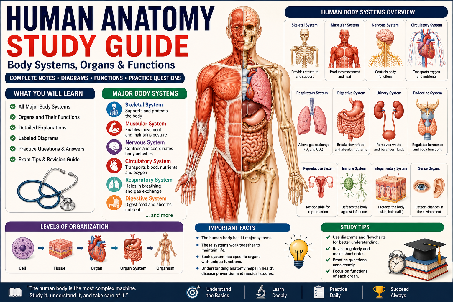
You’ve probably heard the saying that the human body is an incredible machine. It’s one of those phrases that gets repeated so often it starts to lose meaning—until you actually sit down and study anatomy.
When you learn that your heart beats around 100,000 times every single day without you thinking about it, that your kidneys filter about 180 liters of blood daily, or that your nervous system can send signals at speeds of up to 120 meters per second, the word “incredible” suddenly feels like an understatement.
Human anatomy is the study of the body’s structures—what they are, where they are, and how they connect to everything else. It’s the map you need before you can understand how the body functions in health and why things go wrong in disease.
This human anatomy study guide is designed to give you everything in one organized place. Whether you’re a high school student covering body systems for the first time, a college student taking anatomy and physiology, a nursing or medical student building foundational knowledge, or simply someone who wants to understand the body they live in—this guide has you covered.
Every major system is explained clearly. Every important organ is described with its function. Diagrams, vocabulary, practice questions, and exam tips are all included. Let’s build your understanding from the ground up.
Key Takeaways
- Human anatomy studies the structure of the body at levels from cells to the whole organism.
- The body is organized into 11 major organ systems, each with specific structures and functions.
- Systems don’t work in isolation—they’re deeply interconnected and interdependent.
- Understanding anatomy requires learning precise terminology, spatial relationships, and functional context.
- Drawing and labeling diagrams from memory is one of the highest-yield study strategies for anatomy exams.
- This guide covers all 11 organ systems, major organs, sense organs, anatomical terminology, and 35 practice questions across three difficulty levels.
- Real-world applications connect anatomical knowledge to medicine, fitness, nursing, and daily health decisions.
What Is Human Anatomy?
Human anatomy is the scientific study of the structure of the human body—its parts, their arrangement, and their relationships to one another. The word “anatomy” comes from the Greek anatome, meaning “to cut up” or “to dissect,” which reflects how early anatomists learned about the body by carefully examining it.
Anatomy answers the question: What is the body made of, and where is everything located?
It works alongside physiology, which asks: How does each structure function? Together, anatomy and physiology give you a complete picture of the human body. You can’t truly understand one without the other—knowing where the heart is matters much less if you don’t understand why it beats.
Anatomy is studied at different levels:
- Gross anatomy (macroscopic anatomy) — structures visible to the naked eye (organs, bones, muscles)
- Microscopic anatomy (histology) — cell and tissue structures requiring a microscope
- Surface anatomy — external features of the body and their relationship to internal structures
- Regional anatomy — studying all structures in one area (e.g., the thorax or the head)
- Systemic anatomy — studying one organ system at a time across the whole body (the approach used in most courses)
For most high school and introductory college courses, systemic anatomy is the primary approach—and that’s the framework this guide follows.
Why Is Human Anatomy Important?
The practical value of understanding human anatomy stretches across a remarkable range of fields and everyday situations.
In medicine and healthcare, anatomy is the absolute foundation. Surgeons must know the precise location of every structure before making an incision. Nurses need to understand which veins are accessible for IV placement. Radiologists read imaging scans by recognizing normal anatomy and identifying deviations from it. Pharmacists need to understand drug absorption, distribution, and organ-specific effects.
In physical therapy and exercise science, understanding muscle anatomy, joint mechanics, and skeletal structure allows practitioners to design effective rehabilitation programs and prevent injury.
In emergency situations, basic anatomical knowledge helps people respond appropriately. Knowing where to apply pressure to stop bleeding, how CPR works mechanically, or recognizing signs of stroke connects directly to anatomical understanding.
For students, human anatomy is a required foundation for biology, nursing, medicine, physical education, kinesiology, health science, and numerous allied health programs. Performance in these fields often depends significantly on how well you mastered anatomy early.
In everyday life, understanding your own body helps you interpret symptoms, follow medical advice, make informed health decisions, and engage meaningfully with healthcare providers.
History of Human Anatomy
The story of how humans came to understand their own bodies is one of the most fascinating in science—spanning thousands of years, cultural restrictions, revolutionary discoveries, and technological advances.
| Period / Year | Figure / Event | Contribution |
|---|---|---|
| ~3000 BCE | Ancient Egyptians | Practiced embalming; developed early knowledge of organs |
| ~400 BCE | Hippocrates | Described many anatomical structures; laid groundwork for systematic observation |
| ~300 BCE | Herophilus | First systematic human dissections in Alexandria; described brain, nerves, and vessels |
| ~300 BCE | Erasistratus | Distinguished sensory from motor nerves; studied the heart and valves |
| ~150 CE | Galen of Pergamon | Prolific anatomist (mostly animal dissections); his work dominated anatomy for 1,400 years |
| 1316 | Mondino de Luzzi | First systematic anatomy textbook; performed public dissections |
| 1543 | Andreas Vesalius | Published De Humani Corporis Fabrica; corrected Galen’s errors; founded modern anatomy |
| 1628 | William Harvey | Demonstrated the circulation of blood through the body |
| 1661 | Marcello Malpighi | Discovered capillaries using microscopy, completing Harvey’s circulation model |
| 1800s | Various anatomists | Systematic description of muscles, nerves, and organs through cadaveric dissection |
| 1895 | Wilhelm Röntgen | Discovered X-rays, enabling non-invasive visualization of internal anatomy |
| 1970s–present | CT, MRI, ultrasound | Advanced imaging technologies revolutionized clinical and educational anatomy |
| 1994 | Visible Human Project | First complete digital representation of the human body using cross-sectional anatomy |
Understanding this history helps you appreciate that what we know today was earned through careful observation, willingness to question authority, and technological innovation.
Levels of Organization in the Human Body
The human body is organized into a hierarchy of increasing complexity. Understanding this hierarchy is essential because it frames everything else you’ll study in anatomy.
Cells
The cell is the basic structural and functional unit of life. The human body contains approximately 37 trillion cells, each performing specific tasks. There are over 200 distinct cell types in the body—red blood cells, neurons, muscle fibers, epithelial cells, and many more.
Despite their differences, all cells share fundamental features: a cell membrane, cytoplasm, organelles, and (in most cases) a nucleus containing DNA.
Tissues
A tissue is a group of similar cells working together to perform a specific function. The human body has four primary tissue types:
| Tissue Type | Location | Function |
|---|---|---|
| Epithelial | Skin surface, linings of organs, glands | Protection, secretion, absorption, filtration |
| Connective | Bones, blood, fat, cartilage, tendons | Support, connection, transport, energy storage |
| Muscle | Skeletal muscles, heart, digestive organs | Movement, pumping, peristalsis |
| Nervous | Brain, spinal cord, nerves | Signal transmission, coordination, sensation |
Organs
An organ is a structure composed of two or more tissue types working together to perform specific functions. The heart, for example, is made of cardiac muscle tissue, connective tissue, epithelial tissue, and nervous tissue—all working together to pump blood.
The human body has approximately 78 organs, though the exact count depends on how organs are defined.
Organ Systems
An organ system is a group of organs that work together to accomplish a major physiological function. The human body has 11 organ systems:
- Skeletal
- Muscular
- Nervous
- Circulatory (Cardiovascular)
- Respiratory
- Digestive
- Urinary (Excretory)
- Endocrine
- Reproductive
- Immune and Lymphatic
- Integumentary
Organism
The organism is the highest level of organization—the complete, living human being. All systems operate simultaneously and in coordination to maintain life and homeostasis.
Why this hierarchy matters: Every anatomy question you encounter fits somewhere in this hierarchy. Knowing where something sits—whether it’s a cell type, a tissue, an organ, or a system—helps you organize information logically and answer questions more accurately.
Major Human Body Systems Overview
| System | Major Organs/Structures | Primary Function |
|---|---|---|
| Skeletal | Bones, cartilage, ligaments | Support, protection, movement, blood cell production |
| Muscular | Skeletal, cardiac, smooth muscles | Movement, posture, heat generation |
| Nervous | Brain, spinal cord, nerves | Control, coordination, sensation, response |
| Circulatory | Heart, blood vessels, blood | Transport of oxygen, nutrients, and waste |
| Respiratory | Lungs, trachea, bronchi, diaphragm | Gas exchange (O₂ in, CO₂ out) |
| Digestive | Stomach, intestines, liver, pancreas | Digestion and absorption of nutrients |
| Urinary | Kidneys, bladder, ureters, urethra | Waste filtration, fluid and electrolyte balance |
| Endocrine | Glands (pituitary, thyroid, adrenal, etc.) | Hormonal regulation of body functions |
| Reproductive | Gonads, reproductive organs | Reproduction, sex hormone production |
| Immune/Lymphatic | Lymph nodes, spleen, white blood cells | Defense against pathogens, fluid balance |
| Integumentary | Skin, hair, nails, glands | Protection, temperature regulation, sensation |
Skeletal System
The skeletal system is the internal framework of the body. It’s not just a passive scaffold—it’s a dynamic, living system that constantly remodels itself and contributes to multiple physiological processes.
Bones
The adult human skeleton contains 206 bones, though newborns start with approximately 270–300 (many fuse during growth and development).
Bone tissue types:
- Compact bone (cortical bone) — Dense outer layer; provides strength; makes up about 80% of total bone mass
- Spongy bone (cancellous bone) — Lightweight inner layer with a honeycomb structure; found at the ends of long bones; contains red bone marrow
Bone cells:
| Cell Type | Function |
|---|---|
| Osteoblasts | Build new bone tissue |
| Osteoclasts | Break down (resorb) bone tissue |
| Osteocytes | Mature bone cells that maintain bone matrix |
Classification of bones by shape:
| Type | Description | Examples |
|---|---|---|
| Long bones | Longer than wide; cylindrical shaft | Femur, humerus, tibia |
| Short bones | Roughly cube-shaped | Carpals (wrist), tarsals (ankle) |
| Flat bones | Thin and flat; often curved | Skull, sternum, scapula, ribs |
| Irregular bones | Complex shapes | Vertebrae, hip bones, facial bones |
| Sesamoid bones | Small, round, embedded in tendons | Patella (kneecap) |
Key anatomical parts of a long bone:
- Diaphysis — The shaft of the bone
- Epiphysis — The rounded ends
- Periosteum — Outer membrane covering bone surface; contains nerve fibers and blood vessels
- Medullary cavity — Hollow center of the shaft containing yellow bone marrow (fat)
- Endosteum — Inner membrane lining the medullary cavity
- Articular cartilage — Hyaline cartilage covering joint surfaces to reduce friction
Joints
A joint (articulation) is where two or more bones meet. Joints allow varying degrees of movement and provide mechanical stability.
Classification by movement:
| Joint Type | Movement | Examples |
|---|---|---|
| Fibrous (synarthrosis) | No movement | Sutures of the skull |
| Cartilaginous (amphiarthrosis) | Slight movement | Intervertebral discs, pubic symphysis |
| Synovial (diarthrosis) | Free movement | Shoulder, hip, knee, elbow |
Types of synovial joints:
- Hinge — Movement in one plane (flexion/extension): knee, elbow
- Ball and socket — Widest range of movement: hip, shoulder
- Pivot — Rotation: atlas-axis joint (head rotation)
- Saddle — Two directions: thumb carpometacarpal joint
- Gliding (plane) — Sliding movement: carpal bones
- Condyloid — Oval surface allows multiple movements: wrist, knuckles
Functions of the Skeleton
- Support — Provides structural framework that holds the body upright
- Protection — Skull protects the brain; vertebral column protects the spinal cord; rib cage protects heart and lungs
- Movement — Provides attachment points for muscles; bones act as levers
- Mineral storage — Stores calcium (99% of body’s calcium) and phosphorus; released into blood when needed
- Blood cell production (hematopoiesis) — Red bone marrow in flat bones and long bone epiphyses produces red blood cells, white blood cells, and platelets
- Fat storage — Yellow bone marrow stores adipose tissue as an energy reserve
Muscular System
The muscular system is responsible for every movement your body makes—from running to breathing to the beating of your heart.
Types of Muscles
| Muscle Type | Control | Location | Appearance | Characteristics |
|---|---|---|---|---|
| Skeletal muscle | Voluntary | Attached to bones | Striated (striped) | Fast-acting; fatigues |
| Cardiac muscle | Involuntary | Heart wall only | Striated | Never fatigues; self-stimulating |
| Smooth muscle | Involuntary | Walls of organs (gut, bladder, blood vessels) | Non-striated | Slow, sustained contractions |
Major Muscle Groups
Upper body:
- Deltoid — Shoulder abduction; rounded shoulder shape
- Pectoralis major — Chest muscle; arm adduction and flexion
- Trapezius — Upper back; elevates and retracts scapula
- Biceps brachii — Front of upper arm; flexes forearm
- Triceps brachii — Back of upper arm; extends forearm
- Latissimus dorsi — Large back muscle; arm extension and adduction
Core and trunk:
- Rectus abdominis — Front of abdomen; flexes vertebral column (“abs”)
- External obliques — Sides of abdomen; rotation and lateral flexion
- Diaphragm — Dome-shaped muscle separating thorax and abdomen; primary breathing muscle
- Erector spinae — Along the spine; extends and stabilizes the vertebral column
Lower body:
- Quadriceps femoris — Front of thigh (four muscles); extends the knee
- Hamstrings — Back of thigh (three muscles); flexes knee, extends hip
- Gluteus maximus — Buttock; hip extension
- Gastrocnemius — Calf; plantar flexion (pointing foot down)
- Tibialis anterior — Front of lower leg; dorsiflexion (pulling foot up)
- Iliopsoas — Hip flexor; flexes thigh at hip
Functions of the Muscular System
- Movement — Skeletal muscles pull on bones to produce movement at joints
- Posture and stability — Constant low-level muscle contraction maintains upright posture
- Heat production — Muscle contractions generate ~85% of the body’s heat; crucial for temperature regulation
- Circulation support — Skeletal muscle contractions squeeze veins, helping return blood to the heart (the “muscle pump”)
- Protection — Muscles protect underlying organs from mechanical injury
- Respiration — The diaphragm and intercostal muscles power breathing
How muscles contract:
Muscles work on the sliding filament theory. Thin actin filaments slide over thick myosin filaments, shortening the sarcomere (the basic contractile unit). This requires calcium ions (released from the sarcoplasmic reticulum) and ATP. The process is triggered by nerve impulses at the neuromuscular junction.
Antagonistic muscle pairs:
Muscles can only pull, not push. They work in opposing pairs:
- Agonist (prime mover) — produces the desired movement
- Antagonist — relaxes and stretches to allow the movement
- Example: When the biceps contracts (agonist) to flex the elbow, the triceps relaxes (antagonist)
Nervous System
The nervous system is the body’s command and communication network. It detects stimuli, processes information, and coordinates appropriate responses—all at extraordinary speed.
Two main divisions:
- Central Nervous System (CNS) — Brain and spinal cord; processes and integrates information
- Peripheral Nervous System (PNS) — All nerves outside the CNS; carries signals to and from the CNS
Functional divisions of the PNS:
- Somatic nervous system — Controls voluntary skeletal muscle movements
- Autonomic nervous system — Controls involuntary functions (heart rate, digestion, gland secretion)
- Sympathetic division — “Fight or flight” response; increases heart rate, dilates airways, redirects blood to muscles
- Parasympathetic division — “Rest and digest”; slows heart rate, stimulates digestion, conserves energy
Brain
The brain is the most complex organ in the human body. It contains approximately 86 billion neurons and an equal number of glial (support) cells. Protected by the skull and three layers of membranes called meninges, it’s bathed in cerebrospinal fluid (CSF) that cushions it against impacts.
Major regions of the brain:
| Region | Location | Key Functions |
|---|---|---|
| Cerebrum | Largest part; two hemispheres | Conscious thought, voluntary movement, language, memory, sensation |
| Cerebellum | Posterior, below cerebrum | Coordination, balance, fine motor control |
| Brainstem | Connects brain to spinal cord | Controls heart rate, breathing, blood pressure, sleep/wake cycles |
| Hypothalamus | Below thalamus | Regulates temperature, hunger, thirst, sleep, hormone release |
| Thalamus | Central relay station | Routes sensory information to appropriate brain regions |
| Limbic system | Deep within cerebrum | Emotions, memory formation, motivation |
Cerebral lobes and their functions:
| Lobe | Location | Primary Functions |
|---|---|---|
| Frontal | Front of brain | Decision-making, personality, voluntary movement, speech production (Broca’s area) |
| Parietal | Top of brain | Somatosensory processing; spatial awareness; integrating sensory information |
| Temporal | Sides of brain | Hearing, language comprehension (Wernicke’s area), memory, face recognition |
| Occipital | Back of brain | Visual processing |
Spinal Cord
The spinal cord extends from the brainstem to the level of the first or second lumbar vertebra—roughly to your lower back. It’s approximately 45 cm long in adults.
Functions:
- Carries sensory signals up to the brain (ascending tracts)
- Carries motor signals down from the brain (descending tracts)
- Processes reflex arcs independently (without brain involvement)
Reflex arc: A direct pathway that produces a rapid, automatic response without waiting for the brain to process. Example: pulling your hand away from a hot object. The signal goes through the spinal cord and produces a motor response before you’re even consciously aware of pain.
Peripheral Nervous System
The PNS consists of 12 pairs of cranial nerves (connecting directly to the brain) and 31 pairs of spinal nerves (branching from the spinal cord).
Types of PNS neurons:
- Sensory (afferent) neurons — Carry signals FROM sensory receptors TO the CNS
- Motor (efferent) neurons — Carry signals FROM the CNS TO effectors (muscles, glands)
- Interneurons — Connect sensory and motor neurons within the CNS
Neuron structure:
- Dendrites — Branch-like extensions that receive signals
- Cell body (soma) — Contains nucleus; integrates incoming signals
- Axon — Long fiber that carries signals away from the cell body
- Myelin sheath — Fatty insulating layer that speeds signal transmission; produced by Schwann cells (PNS) or oligodendrocytes (CNS)
- Synaptic terminals — Release neurotransmitters to communicate with the next neuron
Circulatory System
The circulatory system—also called the cardiovascular system—is the body’s transport network. It delivers oxygen, nutrients, hormones, and immune cells throughout the body and carries waste products to the appropriate organs for elimination.
Heart
The heart is a hollow, muscular organ about the size of your fist, located slightly left of center in the chest cavity (mediastinum). It beats approximately 60–100 times per minute at rest—over 100,000 times per day.
Heart structure:
| Structure | Description |
|---|---|
| Pericardium | Double-layered sac surrounding the heart; prevents friction |
| Epicardium | Outer layer of the heart wall |
| Myocardium | Thick middle layer of cardiac muscle; the actual pumping tissue |
| Endocardium | Inner layer lining the heart chambers |
| Right atrium | Receives deoxygenated blood from the body via the vena cava |
| Right ventricle | Pumps deoxygenated blood to the lungs via the pulmonary artery |
| Left atrium | Receives oxygenated blood from the lungs via the pulmonary veins |
| Left ventricle | Pumps oxygenated blood to the entire body; thickest wall |
Heart valves (prevent backflow of blood):
- Atrioventricular valves: Tricuspid (right side) and Mitral/Bicuspid (left side) — between atria and ventricles
- Semilunar valves: Pulmonary and Aortic — between ventricles and major arteries
Dual circulation:
- Pulmonary circulation — Right side of heart → Lungs (for gas exchange) → Left side of heart
- Systemic circulation — Left side of heart → Body tissues → Right side of heart
The cardiac cycle:
- Systole — Ventricles contract; blood is ejected into arteries
- Diastole — Ventricles relax and fill with blood
- Heard as “lub-dub” through a stethoscope (closure of AV valves = “lub”; closure of semilunar valves = “dub”)
Cardiac conduction system:
The heart generates its own electrical signals:
- SA node (sinoatrial node) — The pacemaker; initiates each heartbeat in the right atrium
- AV node (atrioventricular node) — Delays signal briefly; allows atria to fully empty before ventricles contract
- Bundle of His — Conducts signal to ventricles
- Purkinje fibers — Distribute signal throughout ventricles, causing simultaneous contraction
Blood
Blood is a specialized connective tissue composed of cells suspended in a liquid matrix called plasma.
Composition of blood:
| Component | Percentage | Description and Function |
|---|---|---|
| Plasma | ~55% | Yellow fluid; carries dissolved nutrients, hormones, proteins, waste products |
| Red blood cells (Erythrocytes) | ~44% | Contain hemoglobin; carry oxygen and some CO₂; no nucleus when mature |
| White blood cells (Leukocytes) | <1% | Immune defense; several types (neutrophils, lymphocytes, monocytes, etc.) |
| Platelets (Thrombocytes) | <1% | Cell fragments; essential for blood clotting (hemostasis) |
Hemoglobin: The iron-containing protein in RBCs that binds oxygen. Each hemoglobin molecule can carry four oxygen molecules. The iron gives blood its red color.
Blood types (ABO system):
Determined by antigens on the surface of RBCs:
- Type A: A antigens
- Type B: B antigens
- Type AB: Both antigens (universal recipient)
- Type O: No antigens (universal donor)
Also important: Rh factor (positive or negative). Rh incompatibility between mother and fetus can cause hemolytic disease of the newborn.
Blood Vessels
Three main types of blood vessels transport blood throughout the body:
| Vessel Type | Direction | Wall Structure | Function |
|---|---|---|---|
| Arteries | Away from heart | Thick, muscular, elastic walls | Carry oxygenated blood under high pressure |
| Capillaries | Connect arteries to veins | Single cell layer (one cell thick) | Site of gas, nutrient, and waste exchange |
| Veins | Toward heart | Thinner walls; contain valves | Return deoxygenated blood to heart under low pressure |
Capillaries are where the actual work of the circulatory system happens. Their walls are just one cell thick—thin enough for oxygen, carbon dioxide, glucose, and other small molecules to diffuse in and out.
Respiratory System
The respiratory system’s primary job is gas exchange—getting oxygen into the blood and carbon dioxide out of it. Without this exchange, cellular respiration would quickly stop, and cells would begin dying within minutes.
Lungs
The lungs are two large, spongy organs filling most of the thoracic (chest) cavity. The right lung has three lobes; the left lung has two (to accommodate the heart). Together they contain approximately 300–500 million alveoli—tiny air sacs where gas exchange occurs.
Lung capacity measurements:
- Tidal volume — Air inhaled/exhaled in a normal breath (~500 mL)
- Vital capacity — Maximum air exhaled after maximum inhalation (~4,500 mL in adult males)
- Residual volume — Air remaining in lungs after maximum exhalation (~1,200 mL)
Pleura: The lungs are covered by a double-layered membrane—the pleura. The space between the layers contains a thin fluid that reduces friction during breathing. Inflammation of the pleura (pleuritis) causes sharp chest pain with breathing.
Airways
The airway is the pathway air travels from the outside world to the alveoli:
Nose/Mouth → Pharynx → Larynx → Trachea → Bronchi → Bronchioles → Alveoli
| Structure | Description |
|---|---|
| Nose/Nasal cavity | Filters, warms, and humidifies incoming air |
| Pharynx | Common passage for air and food; throat region |
| Larynx | Voice box; contains vocal cords; epiglottis prevents food from entering airway |
| Trachea | Windpipe; reinforced by C-shaped cartilage rings; ~12 cm long |
| Bronchi | Two main branches (right and left) entering each lung |
| Bronchioles | Smaller branches; no cartilage; can dilate or constrict |
| Alveoli | Tiny air sacs; site of gas exchange; surrounded by capillaries |
Epiglottis: A flap of cartilage that closes over the glottis (opening to the larynx) during swallowing, directing food into the esophagus and preventing choking. When this fails, food “goes down the wrong way.”
Breathing mechanics:
- Inhalation (inspiration): Diaphragm contracts and flattens; external intercostal muscles contract; chest volume increases; air pressure inside lungs drops below atmospheric pressure; air flows in
- Exhalation (expiration): Diaphragm relaxes and domes upward; chest volume decreases; lung pressure rises above atmospheric; air flows out
This is Boyle’s Law in action: pressure and volume are inversely related.
Gas Exchange
Gas exchange occurs in the alveoli by simple diffusion following concentration gradients:
- Oxygen diffuses from alveoli (high O₂) into blood capillaries (lower O₂)
- Carbon dioxide diffuses from blood capillaries (high CO₂) into alveoli (lower CO₂), where it’s exhaled
Adaptations of alveoli for efficient gas exchange:
- Extremely thin walls (one cell thick) — short diffusion distance
- Enormous total surface area (~70 m²—roughly the size of a tennis court)
- Surrounded by dense capillary network
- Moist surface facilitates gas dissolution
- Coated with surfactant (reduces surface tension; prevents alveoli from collapsing)
Digestive System
The digestive system breaks food down into molecules small enough to be absorbed into the bloodstream and used by cells. This happens through both mechanical digestion (physical breakdown) and chemical digestion (enzyme action).
The alimentary canal (digestive tract):
Mouth → Esophagus → Stomach → Small intestine → Large intestine → Rectum → Anus
Total length of the digestive tract: approximately 9 meters in an adult.
Mouth
Digestion begins in the mouth with both mechanical and chemical processes:
- Teeth mechanically break food into smaller pieces (mastication)
- Tongue mixes food with saliva and pushes it toward the throat
- Salivary glands produce saliva containing:
- Salivary amylase — begins carbohydrate digestion by breaking down starch
- Mucus — lubricates food to form a bolus (ball of food) for swallowing
- Lysozyme — antimicrobial enzyme
Food is formed into a bolus and swallowed through the pharynx into the esophagus.
Esophagus:
The esophagus is a muscular tube (~25 cm) connecting the pharynx to the stomach. It uses peristalsis—rhythmic waves of muscular contraction—to move food downward. The lower esophageal sphincter (cardiac sphincter) prevents stomach acid from flowing back up. When it weakens, the result is acid reflux (GERD).
Stomach
The stomach is a J-shaped muscular organ in the upper left abdomen that temporarily stores food, churns it mechanically, and begins protein digestion.
Stomach functions:
- Stores up to 1–1.5 liters of food
- Mechanical churning converts food to a semi-liquid mass called chyme
- Gastric glands in the stomach wall secrete:
- Hydrochloric acid (HCl) — Creates an acidic environment (pH ~1.5–3.5); kills pathogens; denatures proteins; activates pepsinogen
- Pepsinogen — Converted to active pepsin by HCl; begins protein digestion
- Intrinsic factor — Required for vitamin B12 absorption
- Mucus — Protects stomach lining from acid and enzymes
Food spends 2–4 hours in the stomach before being gradually released into the small intestine through the pyloric sphincter.
Small Intestine
The small intestine is where the majority of chemical digestion and nutrient absorption occurs. Despite its name, it’s approximately 6–7 meters long in a living adult.
Three regions:
- Duodenum — First ~25 cm; receives chyme from stomach; receives bile from the liver and digestive enzymes from the pancreas; most chemical digestion occurs here
- Jejunum — Middle section; primary site of nutrient absorption
- Ileum — Final section; absorbs remaining nutrients, including vitamin B12 and bile salts; connects to large intestine via ileocecal valve
Structural adaptations maximizing absorption:
- Circular folds (plicae circularis) — Large folds of the intestinal wall that increase surface area
- Villi — Finger-like projections of the mucosa; each contains a capillary network and a central lacteal (lymph vessel)
- Microvilli (brush border) — Tiny projections on the surface of each villus; collectively called the brush border; covered with digestive enzymes
These three layers of folding increase the absorptive surface area of the small intestine from ~0.33 m² to approximately 250 m²—roughly the size of a tennis court.
Absorbed nutrients go to:
- Capillaries in villi → Portal vein → Liver (for processing): glucose, amino acids, water-soluble vitamins, minerals
- Lacteals in villi → Lymphatic system → Bloodstream: fats, fat-soluble vitamins (A, D, E, K)
Large Intestine
The large intestine is approximately 1.5 meters long and consists of the cecum, colon (ascending, transverse, descending, sigmoid), rectum, and anal canal.
Functions:
- Absorbs water and electrolytes from undigested material
- Converts liquid chyme to solid or semi-solid feces
- Houses the gut microbiome (trillions of beneficial bacteria)
- Stores feces until defecation
- Bacterial fermentation of undigested material produces some B vitamins and vitamin K
Appendix: A small finger-like projection from the cecum. Its function was once considered vestigial, but current evidence suggests it may play a role in housing beneficial gut bacteria. Appendicitis (inflammation) is a medical emergency requiring surgical removal.
Liver
The liver is the largest internal organ (~1.5 kg) and performs over 500 different functions.
Key functions of the liver:
- Produces bile (stored in the gallbladder; released into the duodenum to emulsify fats)
- Metabolizes carbohydrates, proteins, and fats
- Detoxifies alcohol, drugs, and other harmful substances
- Synthesizes plasma proteins (albumin, clotting factors)
- Stores glycogen, vitamins A, D, B12, and iron
- Converts ammonia (from protein metabolism) to urea (excreted by kidneys)
- Destroys old red blood cells
Pancreas
The pancreas has both endocrine functions (hormonal) and exocrine functions (digestive).
Exocrine functions (digestion):
- Produces pancreatic juice delivered via pancreatic duct to the duodenum
- Contains:
- Pancreatic amylase — digests starch to maltose
- Pancreatic lipase — digests fats to fatty acids and glycerol
- Proteases (trypsinogen, chymotrypsinogen) — digest proteins
- Sodium bicarbonate — neutralizes acidic chyme entering the duodenum
Endocrine functions (hormones):
- Insulin (from beta cells) — Lowers blood glucose; promotes glucose uptake by cells
- Glucagon (from alpha cells) — Raises blood glucose; promotes glycogen breakdown in liver
Urinary System
The urinary system is the body’s filtration and waste management system. Its primary job is to remove metabolic waste products from the blood, regulate fluid and electrolyte balance, and maintain blood pH.
Components: Two kidneys, two ureters, one bladder, one urethra
Kidneys
The kidneys are bean-shaped organs (~11 cm long) located retroperitoneally (behind the peritoneum) on either side of the vertebral column.
The functional unit of the kidney is the nephron. Each kidney contains approximately 1 million nephrons. Each nephron consists of:
- Glomerulus — A knot of capillaries inside Bowman’s capsule where filtration begins
- Bowman’s capsule — Cup-like structure surrounding the glomerulus; collects the filtrate
- Proximal convoluted tubule (PCT) — Reabsorbs ~65% of filtered water, glucose, amino acids, and ions
- Loop of Henle — Creates a concentration gradient in the medulla; important for concentrating urine
- Distal convoluted tubule (DCT) — Further reabsorption and secretion; regulated by hormones
- Collecting duct — Final concentration of urine; regulated by ADH (antidiuretic hormone)
Three processes of urine formation:
- Filtration — Blood is forced under pressure through the glomerulus; small molecules (water, glucose, urea, ions) pass into Bowman’s capsule
- Reabsorption — Useful substances (glucose, amino acids, most water) are transported back into the blood
- Secretion — Additional waste products (H⁺ ions, drugs) are actively transported from blood into the tubule
Hormonal control of kidney function:
- ADH (antidiuretic hormone) — Released by posterior pituitary; increases water reabsorption in collecting ducts; concentrated urine
- Aldosterone — Released by adrenal cortex; increases Na⁺ reabsorption (water follows); raises blood pressure
- Renin-angiotensin-aldosterone system (RAAS) — Activated when blood pressure drops; complex cascade leading to blood pressure restoration
Other functions of the kidneys:
- Regulate blood pH by excreting H⁺ and reabsorbing bicarbonate
- Produce erythropoietin (EPO)—stimulates RBC production
- Activate vitamin D
Endocrine System
The endocrine system communicates through hormones—chemical messengers secreted directly into the bloodstream by glands, traveling to target cells that have specific receptors for each hormone.
Unlike the nervous system (fast, short-duration signals), the endocrine system acts more slowly but produces longer-lasting effects.
Major endocrine glands and hormones:
| Gland | Location | Key Hormones | Functions |
|---|---|---|---|
| Hypothalamus | Brain | Releasing/inhibiting hormones | Controls pituitary gland; regulates temperature, hunger, thirst |
| Pituitary gland (anterior) | Below hypothalamus | GH, TSH, ACTH, FSH, LH, Prolactin | Master gland; controls other glands |
| Pituitary gland (posterior) | Below hypothalamus | ADH, Oxytocin | Water balance; uterine contractions; bonding |
| Thyroid | Neck | T3, T4 (thyroid hormones), Calcitonin | Metabolism rate; calcium regulation |
| Parathyroid | Behind thyroid | PTH (parathyroid hormone) | Raises blood calcium |
| Adrenal cortex | Above kidneys | Cortisol, Aldosterone, Androgens | Stress response; Na⁺/K⁺ balance; sex hormones |
| Adrenal medulla | Above kidneys | Adrenaline (epinephrine), Noradrenaline | Fight-or-flight response |
| Pancreas (islets) | Abdomen | Insulin, Glucagon | Blood glucose regulation |
| Gonads (testes) | Scrotum | Testosterone | Male sex characteristics; sperm production |
| Gonads (ovaries) | Pelvis | Estrogen, Progesterone | Female sex characteristics; menstrual cycle |
| Pineal gland | Brain | Melatonin | Sleep-wake cycles; circadian rhythm |
| Thymus | Chest | Thymosin | T-lymphocyte maturation (immune function) |
Feedback mechanisms:
Most hormone release is controlled by negative feedback—when hormone levels reach a certain point, they signal the gland to stop producing more. Example: High thyroid hormone levels signal the pituitary to stop releasing TSH, which reduces thyroid hormone production.
Positive feedback amplifies a response (less common): Example: Oxytocin during childbirth intensifies contractions until delivery.
Reproductive System
The reproductive system’s primary function is to produce offspring. It also produces sex hormones that influence a wide range of physiological processes.
Male Reproductive System:
| Structure | Function |
|---|---|
| Testes | Produce sperm (spermatogenesis) and testosterone |
| Epididymis | Sperm maturation and storage |
| Vas deferens | Tube transporting sperm from epididymis to urethra |
| Seminal vesicles | Produce fructose-rich fluid nourishing sperm |
| Prostate gland | Produces alkaline fluid protecting sperm in the acidic vagina |
| Bulbourethral glands | Produce lubricating fluid (pre-ejaculate) |
| Urethra | Carries semen and urine out of the body (not simultaneously) |
| Penis | Organ of copulation and urination |
Spermatogenesis: Production of sperm cells in the seminiferous tubules of the testes through meiosis. Takes approximately 74 days. Temperature-sensitive—the testes are in the scrotum because sperm production requires a temperature ~2–3°C below core body temperature.
Female Reproductive System:
| Structure | Function |
|---|---|
| Ovaries | Produce oocytes (eggs) and sex hormones (estrogen, progesterone) |
| Fallopian tubes (oviducts) | Carry eggs from ovary to uterus; site of fertilization |
| Uterus | Site of fetal development; muscular wall (myometrium) |
| Cervix | Lower portion of uterus; opens during childbirth |
| Vagina | Birth canal; receives penis during copulation |
| Vulva | External genitalia |
Menstrual cycle (~28 days):
- Menstruation (days 1–5) — Shedding of uterine lining
- Follicular phase (days 1–13) — FSH stimulates follicle development; rising estrogen thickens uterine lining
- Ovulation (day 14) — LH surge triggers release of egg from ovary
- Luteal phase (days 15–28) — Corpus luteum produces progesterone; prepares uterus for implantation; if no fertilization, corpus luteum degenerates and cycle repeats
Immune and Lymphatic System
The immune system is the body’s defense force. The lymphatic system works alongside it, maintaining fluid balance and transporting immune cells.
Two lines of immune defense:
1. Innate immunity (non-specific):
- First line of defense: Physical barriers (skin, mucous membranes, cilia)
- Second line: Internal non-specific responses (fever, inflammation, phagocytes, natural killer cells, complement proteins)
- Responds immediately; doesn’t distinguish between specific pathogens
2. Adaptive immunity (specific):
- Third line of defense: Specific responses to identified antigens
- Humoral immunity: B lymphocytes produce antibodies targeting specific antigens
- Cell-mediated immunity: T lymphocytes directly attack infected or cancerous cells
- Creates immunological memory — the basis of vaccination
Key immune cells:
| Cell Type | Function |
|---|---|
| Neutrophils | Most abundant WBC; engulf and destroy bacteria (phagocytosis) |
| Macrophages | Engulf pathogens and cellular debris; present antigens to T cells |
| Natural killer (NK) cells | Kill virus-infected cells and cancer cells |
| B lymphocytes | Produce antibodies; differentiate into plasma cells |
| T helper cells (CD4+) | Activate other immune cells; coordinate immune response |
| T cytotoxic cells (CD8+) | Kill infected or abnormal cells directly |
| Memory cells | Long-lived; enable rapid response to previously encountered antigens |
Lymphatic system structures:
- Lymph nodes — Filter lymph fluid; sites of immune cell activation
- Spleen — Filters blood; destroys old red blood cells; houses immune cells
- Thymus — Matures T lymphocytes
- Tonsils — Guard against pathogens entering through mouth and nose
- Lymphatic vessels — Collect excess interstitial fluid and return it to bloodstream
Integumentary System (Skin)
The integumentary system includes the skin and its appendages—hair, nails, and glands. Skin is the largest organ of the human body, covering approximately 1.5–2 m² of surface area.
Three layers of skin:
| Layer | Description | Key Structures |
|---|---|---|
| Epidermis | Outermost layer; avascular; waterproof | Keratinocytes, melanocytes (produce melanin), Langerhans cells (immune), stratum corneum |
| Dermis | Middle layer; thick; contains blood vessels and nerves | Collagen, elastin, hair follicles, sweat glands, sebaceous glands, sensory receptors |
| Hypodermis (subcutaneous) | Deepest layer; not technically part of skin | Adipose tissue, larger blood vessels; connects skin to underlying structures |
Functions of the integumentary system:
- Protection — Physical barrier against pathogens, UV radiation, dehydration, and mechanical injury
- Temperature regulation — Sweat glands cool the body; blood vessel dilation/constriction adjusts heat loss
- Sensation — Contains receptors for touch, pressure, pain, vibration, and temperature
- Vitamin D synthesis — Skin converts UV radiation to vitamin D3, which is then activated by the liver and kidneys
- Excretion — Sweat removes small amounts of waste (urea, salts)
- Immunity — Langerhans cells in the epidermis present antigens to immune cells
Sense Organs
The sense organs are specialized structures that detect stimuli from the environment and transmit sensory information to the brain via sensory nerves.
Eyes
The eyes detect light and convert it into nerve impulses the brain interprets as vision.
Key structures of the eye:
| Structure | Function |
|---|---|
| Cornea | Transparent outer covering; refracts light entering the eye |
| Iris | Colored part; controls pupil size (regulates light entry) |
| Pupil | Opening through which light enters |
| Lens | Focuses light onto the retina; shape changed by ciliary muscles (accommodation) |
| Retina | Light-sensitive layer containing photoreceptors |
| Rods | Photoreceptors for dim light; peripheral vision; no color |
| Cones | Photoreceptors for bright light and color; concentrated at fovea |
| Fovea centralis | Area of sharpest vision; highest cone density |
| Optic nerve | Carries visual signals from retina to brain (occipital lobe) |
| Vitreous humor | Gel-like fluid filling posterior chamber; maintains eye shape |
| Aqueous humor | Watery fluid in anterior chamber; provides nutrients; maintains intraocular pressure |
Ears
The ears serve two functions: hearing and balance (vestibular function).
Three regions of the ear:
| Region | Structures | Function |
|---|---|---|
| Outer ear | Pinna, ear canal, tympanic membrane | Collects and funnels sound; tympanic membrane vibrates with sound waves |
| Middle ear | Malleus, incus, stapes (ossicles); Eustachian tube | Amplifies vibrations; Eustachian tube equalizes pressure |
| Inner ear | Cochlea, semicircular canals, vestibule | Cochlea: converts vibrations to nerve signals (hearing); Semicircular canals: detect rotation (balance) |
Nose
The nose is the primary organ for olfaction (smell) and also conditions incoming air.
- The nasal cavity is lined with olfactory epithelium containing olfactory receptor cells
- Chemicals dissolve in the mucus layer and bind to receptor proteins on olfactory neurons
- Signals travel via the olfactory nerve directly to the olfactory bulb in the brain (closest sensory pathway to the limbic system—which explains why smells trigger strong memories and emotions)
- The nose also warms, filters, and humidifies air before it reaches the lungs
Tongue
The tongue is the primary organ of taste (gustation).
- Covered with papillae (small projections), some of which contain taste buds
- Taste buds contain gustatory cells that detect dissolved chemicals in saliva
- Five primary tastes: Sweet, sour, salty, bitter, umami (savory)
- Taste signals travel via cranial nerves to the taste cortex in the brain
- What we experience as “flavor” is a combination of taste and smell—which is why food tastes bland when you have a blocked nose
Skin (as a Sense Organ)
The skin contains multiple types of sensory receptors:
| Receptor | Sensation Detected |
|---|---|
| Meissner’s corpuscles | Light touch, texture |
| Pacinian corpuscles | Deep pressure, vibration |
| Merkel’s discs | Fine touch, edges, texture |
| Ruffini endings | Skin stretch, sustained pressure |
| Free nerve endings | Pain, temperature, itch |
Human Body Organs and Their Functions (Comparison Table)
| Organ | System | Primary Function |
|---|---|---|
| Brain | Nervous | Control center; processes all sensory and motor information |
| Heart | Circulatory | Pumps blood throughout the body |
| Lungs | Respiratory | Gas exchange (O₂/CO₂) |
| Liver | Digestive/Metabolic | Detoxification; bile production; metabolic regulation |
| Kidneys | Urinary | Filter blood; produce urine; regulate fluid balance |
| Stomach | Digestive | Mechanical and chemical digestion of food |
| Small intestine | Digestive | Nutrient absorption |
| Large intestine | Digestive | Water absorption; waste processing |
| Pancreas | Digestive/Endocrine | Digestive enzymes; insulin and glucagon |
| Spleen | Immune/Lymphatic | Filters blood; immune cell production |
| Thyroid | Endocrine | Regulates metabolism; calcium balance |
| Skin | Integumentary | Protection; temperature regulation; sensation |
| Muscles | Muscular | Movement; heat production |
| Bones | Skeletal | Support; protection; blood cell production |
| Ovaries/Testes | Reproductive | Gamete and sex hormone production |
Organ Systems and Their Main Functions (Comparison Table)
| Organ System | Primary Function | Key Organs |
|---|---|---|
| Skeletal | Structure, support, protection | Bones, cartilage, ligaments |
| Muscular | Movement, posture, heat | Skeletal, cardiac, smooth muscles |
| Nervous | Control, communication, sensation | Brain, spinal cord, nerves |
| Circulatory | Transport of substances | Heart, blood vessels, blood |
| Respiratory | Gas exchange | Lungs, trachea, diaphragm |
| Digestive | Nutrient processing and absorption | Stomach, intestines, liver, pancreas |
| Urinary | Waste removal, fluid balance | Kidneys, bladder, ureters |
| Endocrine | Hormonal regulation | Pituitary, thyroid, adrenal, pancreas |
| Reproductive | Reproduction, sex hormones | Gonads, uterus, penis |
| Immune/Lymphatic | Defense, fluid return | Lymph nodes, spleen, WBCs |
| Integumentary | Protection, temperature, sensation | Skin, hair, nails |
Human Anatomy Terminology Every Student Should Know
Anatomical terminology provides a universal language so that descriptions are clear regardless of body position.
Anatomical position: Body standing upright, facing forward, arms at sides with palms facing forward. All descriptions assume this position.
Directional terms:
| Term | Meaning | Example |
|---|---|---|
| Superior | Above | The head is superior to the shoulders |
| Inferior | Below | The stomach is inferior to the lungs |
| Anterior (ventral) | Toward the front | The sternum is anterior to the heart |
| Posterior (dorsal) | Toward the back | The spine is posterior to the stomach |
| Medial | Toward the midline | The nose is medial to the eyes |
| Lateral | Away from the midline | The ears are lateral to the nose |
| Proximal | Closer to the point of attachment | The elbow is proximal to the wrist |
| Distal | Further from the point of attachment | The fingers are distal to the wrist |
| Superficial | Closer to the body surface | The skin is superficial to the muscles |
| Deep | Further from the body surface | The bones are deep to the muscles |
| Ipsilateral | Same side | The right lung and right kidney are ipsilateral |
| Contralateral | Opposite sides | Left eye and right hand are contralateral |
Body planes:
| Plane | Description | Divides Body Into |
|---|---|---|
| Sagittal | Vertical plane running front to back | Left and right sections |
| Midsagittal (median) | Sagittal plane through exact midline | Equal left and right halves |
| Coronal (frontal) | Vertical plane running side to side | Anterior and posterior sections |
| Transverse (horizontal) | Horizontal plane | Superior and inferior sections |
Body cavities:
| Cavity | Location | Contents |
|---|---|---|
| Cranial | Skull | Brain |
| Vertebral (spinal) | Vertebral column | Spinal cord |
| Thoracic | Chest | Heart, lungs, esophagus, trachea |
| Abdominal | Below diaphragm | Digestive organs, liver, spleen, kidneys |
| Pelvic | Below abdomen | Bladder, reproductive organs, rectum |
Movement terms:
- Flexion — Decreasing the angle at a joint (bending)
- Extension — Increasing the angle at a joint (straightening)
- Abduction — Moving a limb away from the midline
- Adduction — Moving a limb toward the midline
- Rotation — Turning around a long axis
- Circumduction — Circular movement combining flexion, extension, abduction, adduction
Common Human Anatomy Diagrams to Practice
Drawing diagrams from memory is one of the most effective ways to study anatomy. These are the most important:
1. The human skeleton (anterior and posterior views)
Label: skull, mandible, clavicle, sternum, ribs, humerus, radius, ulna, carpals, femur, tibia, fibula, tarsals, vertebral column, pelvis, patella, scapula
2. The heart (cross-sectional view)
Label: all four chambers, four valves, aorta, pulmonary artery, pulmonary veins, superior and inferior vena cava, myocardium, pericardium, SA node, AV node
3. The brain (lateral view and midsagittal view)
Label: four cerebral lobes, cerebellum, brainstem (midbrain, pons, medulla), hypothalamus, thalamus, corpus callosum
4. The nephron
Label: glomerulus, Bowman’s capsule, PCT, loop of Henle (descending and ascending), DCT, collecting duct, afferent and efferent arterioles
5. The digestive system
Label: mouth, esophagus, stomach, small intestine (duodenum, jejunum, ileum), large intestine, rectum, anus, liver, gallbladder, pancreas
6. The respiratory system
Label: nasal cavity, pharynx, larynx, trachea, primary bronchi, secondary bronchi, bronchioles, alveoli, diaphragm, pleura
7. The eye (cross-section)
Label: cornea, iris, pupil, lens, retina, fovea, optic nerve, sclera, vitreous humor, aqueous humor, ciliary muscles
8. The ear (cross-section)
Label: pinna, ear canal, tympanic membrane, malleus, incus, stapes, cochlea, Eustachian tube, semicircular canals, auditory nerve
9. A synovial joint (e.g., the knee)
Label: articular cartilage, synovial membrane, synovial fluid, joint capsule, ligaments, menisci (for the knee)
10. The neuron
Label: dendrites, cell body, nucleus, axon, myelin sheath, nodes of Ranvier, synaptic terminals
Real-Life Applications of Human Anatomy
Understanding anatomy isn’t just for exams—it connects directly to countless real-world situations.
Medical diagnosis and treatment:
When you describe symptoms to a doctor, anatomical knowledge helps locate the problem. Right lower quadrant pain suggests appendicitis; left-sided chest pain radiating to the arm suggests cardiac involvement. Doctors cannot diagnose or treat without anatomical literacy.
Exercise and fitness:
Personal trainers, coaches, and athletes use anatomical knowledge to design effective training programs, understand injury mechanisms, and optimize movement patterns. Knowing that the quadriceps extend the knee helps design exercises targeting that muscle group.
First aid and emergency response:
CPR compresses the heart between the sternum and vertebral column. Knowing the femoral artery’s location helps apply effective pressure for severe leg bleeding. The Heimlich maneuver dislodges foreign objects by understanding the anatomy of the airway.
Nutrition and digestion:
Understanding the digestive system helps explain why certain dietary choices matter—why fiber supports bowel health, why fat-soluble vitamins need dietary fat for absorption, and why people with certain digestive disorders need specific dietary modifications.
Drug administration:
Different administration routes exploit different anatomical pathways. Oral medications pass through the digestive system. Intravenous (IV) medications enter the bloodstream directly. Inhalants reach the lungs through the respiratory tract. Topical medications penetrate through skin layers.
Maternal and child health:
Understanding reproductive anatomy, fetal development, and the mechanics of childbirth helps healthcare providers support pregnancy and delivery safely.
Common Mistakes Students Make
Mistake 1: Learning systems in total isolation
The most common error. The body’s systems are deeply interconnected. The circulatory system delivers oxygen that the respiratory system collects. The kidneys filter blood produced in red bone marrow. The endocrine system regulates digestive function. Always ask: how does this system connect to others?
Mistake 2: Memorizing organ names without understanding functions
Knowing that the liver is in the right upper quadrant is useless without understanding what it does. Always pair structure with function—and understand why the structure enables the function.
Mistake 3: Mixing up directional terms
Superior/inferior, proximal/distal, medial/lateral, anterior/posterior—these get confused regularly, especially under exam pressure. Practice using them in actual sentences about real body structures until they’re automatic.
Mistake 4: Confusing the pulmonary and systemic circuits
Students regularly mix up which side of the heart handles oxygenated versus deoxygenated blood. Remember: the right side receives deoxygenated blood from the body and sends it to the lungs. The left side receives oxygenated blood from the lungs and sends it to the body.
Mistake 5: Forgetting to label diagrams completely
An unlabeled or partially labeled diagram scores poorly. Practice drawing full, labeled diagrams. Every structure on the diagram needs a label, and every label needs to be accurate.
Mistake 6: Confusing smooth, skeletal, and cardiac muscle
These are three distinct tissue types with different structures, locations, and control mechanisms. Know all three—don’t blur the distinctions.
Mistake 7: Underestimating the endocrine system
Students often focus heavily on systems like the circulatory and nervous systems and underprep the endocrine system. Hormone functions are heavily tested at AP, IB, and college levels.
Mistake 8: Neglecting the connections between anatomy and disease
Exams increasingly ask application questions—explaining why a blocked coronary artery causes a heart attack, or how kidney failure affects blood pressure. You need to be able to apply anatomical knowledge to clinical scenarios, not just recite facts.
Best Tips to Study Human Anatomy Faster
1. Learn using the structure-function principle
Never just memorize what something is. Always ask why it’s built that way. The folded inner membrane of mitochondria maximizes surface area for ATP production. The alveoli have thin walls to minimize diffusion distance. Structure and function are inseparable in anatomy.
2. Use layered diagram drawing
Start with a basic body outline. Add one system at a time. This builds spatial understanding that text alone can’t provide. Start with the skeleton, then add muscles, then organs.
3. Create a system-by-system comparison chart
Make a table: System | Major Organs | Input | Process | Output | Connects to. Filling this out for all 11 systems gives you a bird’s-eye view of the entire body.
4. Use the “trace the path” technique
For any substance or signal, trace it from start to finish. Trace a molecule of oxygen from outside air to a muscle cell. Trace a glucose molecule from food to ATP. Trace a nerve signal from a touch receptor to the brain. Following these journeys reveals how systems connect.
5. Watch anatomy videos and animations
Anatomy is inherently spatial. Text alone often isn’t enough for structures like the heart, the ear, or the nephron. High-quality animations make these structures comprehensible in minutes.
6. Use spaced repetition for terminology
Anatomical terminology is vocabulary-heavy. Use flashcard apps with spaced repetition (like Anki) to review terms daily over weeks rather than cramming them the night before an exam.
7. Teach it out loud
Explain a body system as if teaching it to someone who knows nothing about biology. When you stumble, you’ve found a gap. When it flows, you know it.
8. Practice clinical application questions
Don’t just study structures—practice applying them. “A patient has a blocked left coronary artery. Which heart tissue will be most immediately affected?” These application questions are common in nursing, medical, and advanced biology exams.
Human Anatomy Practice Questions
20 Multiple Choice Questions (MCQs)
1. Which bone is found in the upper arm?
- A) Femur B) Radius C) Humerus D) Ulna
2. What is the function of red bone marrow?
- A) Fat storage B) Calcium storage C) Blood cell production D) Hormone production
3. Which type of muscle is found in the wall of the heart?
- A) Skeletal B) Cardiac C) Smooth D) Voluntary
4. What is the primary function of the alveoli?
- A) Filter air B) Gas exchange C) Warm incoming air D) Produce mucus
5. Which chamber of the heart pumps blood to the entire body?
- A) Right atrium B) Right ventricle C) Left atrium D) Left ventricle
6. What is the name of the pacemaker of the heart?
- A) SA node B) AV node C) Bundle of His D) Purkinje fibers
7. Where does the majority of nutrient absorption occur?
- A) Stomach B) Large intestine C) Small intestine D) Esophagus
8. What is the functional unit of the kidney?
- A) Glomerulus B) Loop of Henle C) Nephron D) Bowman’s capsule
9. Which lobe of the brain is responsible for vision?
- A) Frontal B) Parietal C) Temporal D) Occipital
10. What term describes the movement of a limb away from the body’s midline?
- A) Adduction B) Abduction C) Flexion D) Extension
11. Which hormone is responsible for regulating blood glucose by lowering it?
- A) Glucagon B) Cortisol C) Insulin D) Adrenaline
12. What prevents food from entering the trachea during swallowing?
- A) Larynx B) Trachea C) Epiglottis D) Uvula
13. Which cells in the eye are responsible for color vision?
- A) Cones B) Rods C) Ganglia D) Bipolar cells
14. What is the anatomical term for the front of the body?
- A) Posterior B) Superior C) Anterior D) Medial
15. Which blood vessel carries oxygenated blood away from the heart to the body?
- A) Pulmonary artery B) Vena cava C) Pulmonary vein D) Aorta
16. Where is the cochlea located?
- A) Outer ear B) Middle ear C) Inner ear D) Nasal cavity
17. Which organ produces bile?
- A) Gallbladder B) Pancreas C) Liver D) Stomach
18. What type of joint is the knee?
- A) Hinge B) Ball and socket C) Pivot D) Saddle
19. Which hormone is released during a “fight or flight” response?
- A) Insulin B) Oxytocin C) Adrenaline (epinephrine) D) Melatonin
20. What is the term for the fluid-filled sac surrounding the heart?
- A) Pleura B) Peritoneum C) Pericardium D) Endocardium
10 Short Answer Questions
1. Describe the path of a red blood cell from the right atrium through the pulmonary circulation and back to the left ventricle. Name all chambers, valves, and major vessels it passes through.
2. Explain how the structure of the small intestine is adapted to maximize nutrient absorption. Include at least three specific structural features in your answer.
3. Describe the nervous system’s response to placing your hand on a hot surface. Distinguish between the reflex response and the conscious perception of pain, explaining why the reflex happens faster.
4. Explain the three processes involved in urine formation in the nephron. State where each process occurs and give one specific example of a substance involved in each process.
5. Compare the roles of the sympathetic and parasympathetic divisions of the autonomic nervous system. Give two specific examples of how each division affects target organs differently.
6. Describe the structure and function of the three types of muscle tissue. Include whether each is voluntary or involuntary and give a specific location for each.
7. Explain how the body maintains blood glucose homeostasis after a high-carbohydrate meal. Include the role of the pancreas, insulin, and the liver. What would happen if this mechanism failed?
8. Describe how sound waves are converted into nerve impulses by the ear. Trace the path from the outer ear to the brain, naming the key structures involved at each stage.
9. Explain the role of the liver in digestion. Name at least five distinct functions the liver performs and explain why liver disease can have widespread effects on the body.
10. Describe the innate and adaptive immune responses to a bacterial infection. Explain how B lymphocytes contribute to immunity and why vaccination is effective based on this mechanism.
5 Long Answer Questions
1. Describe the structure and function of the cardiovascular system in detail. Explain the cardiac cycle, the conduction system of the heart, the role of blood vessels, and the composition of blood. Explain how the cardiovascular system works with the respiratory system to deliver oxygen to all body cells, and discuss what happens physiologically during a myocardial infarction (heart attack). (15 marks)
2. Explain how the human body maintains homeostasis, using three specific examples from different organ systems. For each example, describe the stimulus, the control center, the effector, and the response. Include one example of negative feedback and explain why most homeostatic mechanisms use this type of feedback. (15 marks)
3. Compare and contrast the skeletal, muscular, and nervous systems in terms of their structure, function, and how they work together to produce voluntary movement. Use the example of picking up a cup of water to trace the entire process from sensory input through neural processing to muscular contraction and skeletal movement. (15 marks)
4. Describe the digestive system as an integrated processing pathway. For each major region from the mouth to the large intestine, describe the mechanical and chemical processes occurring, the enzymes or secretions involved, and what nutrients or substances are absorbed at each stage. Include the role of the liver and pancreas as accessory organs. (15 marks)
5. Discuss the endocrine system and its role in regulating major body functions. Choose five hormones, describe where they are produced, what triggers their release, their target organs, and their effects. Explain the concept of feedback regulation using the hypothalamus-pituitary axis as your primary example and discuss what happens when hormonal regulation fails, using one specific endocrine disorder as your example. (15 marks)
Human Anatomy Revision Checklist
Work through each item carefully. Only check it off when you can explain or demonstrate it confidently.
Foundations
- Explain what human anatomy studies and how it differs from physiology
- Describe the five levels of organization from cell to organism
- Name all four primary tissue types and their locations and functions
- List all 11 organ systems with their primary functions
Anatomical Language
- Define all directional terms (superior, inferior, medial, lateral, etc.)
- Describe the three main body planes (sagittal, coronal, transverse)
- Name all major body cavities and their contents
- Define all movement terms (flexion, extension, abduction, adduction, etc.)
Skeletal System
- State the number of bones in the adult human skeleton
- Classify bones by shape with examples of each
- Label a long bone diagram with all major parts
- Name and describe the three types of joints
- List all six functions of the skeletal system
Muscular System
- Compare the three types of muscle tissue in a table
- Name and locate major muscle groups of the upper and lower body
- Explain the sliding filament theory of muscle contraction
- Describe the concept of antagonistic muscle pairs with an example
Nervous System
- Draw and label a neuron with all major structures
- Describe the structure and functions of the brain’s major regions
- Distinguish between the CNS and PNS
- Compare sympathetic and parasympathetic divisions
- Explain how a reflex arc works
Circulatory System
- Draw and label a diagram of the heart
- Trace blood flow through both pulmonary and systemic circuits
- Explain the cardiac conduction system
- Compare arteries, veins, and capillaries
- Describe the composition and functions of blood
Respiratory System
- Trace the path of air from nose to alveoli
- Explain the mechanics of inhalation and exhalation
- Describe how gas exchange occurs in the alveoli
- Explain why alveoli are adapted for efficient gas exchange
Digestive System
- Trace food from mouth to anus with processes at each stage
- Describe the role of each accessory organ (liver, pancreas, gallbladder)
- Explain how the small intestine is structured for maximum absorption
- Describe the functions of the large intestine
Urinary System
- Draw and label a nephron
- Explain the three processes of urine formation
- Describe how ADH and aldosterone regulate kidney function
Endocrine System
- Name all major endocrine glands and their hormones
- Explain negative and positive feedback with examples
- Describe blood glucose regulation by insulin and glucagon
Other Systems
- Describe the layers and functions of the skin
- Explain the innate and adaptive immune responses
- Name the structures and functions of the eye and ear
- Describe the male and female reproductive systems
Best Books for Human Anatomy
| Book | Best For | Level |
|---|---|---|
| Gray’s Anatomy for Students – Drake et al. | Comprehensive reference and clinical applications | Medical / University |
| Human Anatomy & Physiology – Marieb & Hoehn | Gold standard for introductory A&P courses | College introductory |
| Principles of Anatomy and Physiology – Tortora & Derrickson | Detailed and well-illustrated; widely used | College |
| Atlas of Human Anatomy – Frank Netter | Visual atlas; the best anatomical illustrations available | Medical / University |
| Clinical Anatomy Made Ridiculously Simple – Goldberg | Fast, accessible clinical anatomy | Medical students |
| Campbell Biology – Urry et al. | Body systems coverage within broader biology | AP / College |
| Oxford IB Biology Course Companion | IB-specific anatomy and physiology content | IB |
| CGP GCSE Biology | UK GCSE body systems revision | GCSE |
| Cracking the AP Biology Exam – Princeton Review | AP-focused body systems review | AP |
Free Online Human Anatomy Resources
1. Khan Academy — Human Anatomy & Physiology
khanacademy.org — Free videos and exercises covering all major body systems. Clearly explained, well-organized, and completely free.
2. Visible Body
visiblebody.com — Interactive 3D anatomy models allowing you to rotate, isolate, and explore every body structure. Some free content; widely used by students and healthcare professionals.
3. GetBodySmart
getbodysmart.com — Animated tutorials and quizzes covering all organ systems. Particularly effective for visual learners. Free and regularly updated.
4. Innerbody Research
innerbody.com — Detailed interactive anatomy guides for every body system. Well-written explanations alongside 3D visuals.
5. OpenStax Anatomy and Physiology (Free Textbook)
openstax.org — A complete, peer-reviewed anatomy and physiology textbook freely available online or as a PDF download. Widely used in college courses as a primary or supplementary text.
Frequently Asked Questions
1. What is the difference between anatomy and physiology?
Anatomy studies the structure of the body—what parts exist, where they’re located, and how they’re arranged. Physiology studies how those structures function—what they do and how they work. The two are inseparable: you can’t fully understand function without structure, and structure only makes sense in light of function. Most courses teach them together as “anatomy and physiology.”
2. How many organs does the human body have?
The human body has approximately 78 recognized organs, though the exact number depends on how “organ” is defined. The five vital organs—without which life cannot be sustained—are the brain, heart, lungs, liver, and kidneys.
3. What is the largest organ in the human body?
The skin (part of the integumentary system) is the largest organ, covering approximately 1.5–2 m² of surface area and weighing about 3.5–4 kg in an average adult. If we’re talking about internal organs, the liver is the largest.
4. How does the circulatory system work with the respiratory system?
They work as an integrated gas exchange team. The respiratory system brings oxygen into the alveoli and expels carbon dioxide. The circulatory system sends deoxygenated blood to the lungs via the pulmonary artery, picks up oxygen, and delivers oxygenated blood to all body tissues via the aorta. At the tissues, it drops off oxygen and picks up carbon dioxide, which is then carried back to the lungs. Neither system can function effectively without the other.
5. What is homeostasis and how does the body maintain it?
Homeostasis is the maintenance of a stable internal environment despite external changes. The body regulates temperature, blood glucose, blood pH, fluid balance, and many other variables within narrow ranges. It does this through negative feedback loops: a sensor detects a change, a control center (often the brain or an endocrine gland) processes the signal, and an effector (muscle or gland) responds to restore balance.
6. What is the difference between the central and peripheral nervous systems?
The central nervous system (CNS) consists of the brain and spinal cord—it processes and integrates all information and coordinates responses. The peripheral nervous system (PNS) includes all the nerves outside the CNS—it carries sensory signals to the CNS and motor signals from the CNS to muscles and glands. The PNS is the body’s communication network; the CNS is the processing center.
7. What is the difference between voluntary and involuntary muscles?
Voluntary muscles (skeletal muscles) are under conscious control—you decide to move them. Involuntary muscles (cardiac and smooth muscles) operate independently of conscious control. The heart beats, the digestive tract undergoes peristalsis, and blood vessels change diameter without you having to think about it.
8. How does the immune system protect the body?
The immune system uses multiple layers of defense. Physical barriers (skin, mucus, cilia) are the first line. If pathogens get past these, innate immune responses (phagocytes, inflammation, fever) activate immediately. If the infection persists, the adaptive immune system produces specific antibodies (from B cells) and activates cytotoxic T cells to destroy infected cells. Memory cells from the adaptive response allow faster, stronger responses to future encounters with the same pathogen—which is the principle behind vaccination.
9. What is the role of the liver in the body?
The liver is one of the most multifunctional organs in the body, performing over 500 functions. Key roles include: producing bile for fat digestion, metabolizing carbohydrates, proteins, and fats, detoxifying alcohol and drugs, synthesizing blood clotting factors and plasma proteins, storing glycogen and vitamins, converting ammonia to urea, and destroying old red blood cells. Because of this diversity, liver disease has widespread effects on the entire body.
10. What is the difference between arteries, veins, and capillaries?
Arteries carry blood away from the heart under high pressure—they have thick, muscular, elastic walls to handle this pressure. Most arteries carry oxygenated blood (except the pulmonary arteries). Veins carry blood toward the heart under low pressure—they have thinner walls and internal valves that prevent backflow. Most veins carry deoxygenated blood (except the pulmonary veins). Capillaries are microscopic vessels one cell thick, connecting arteries to veins—they’re where the actual exchange of gases, nutrients, and waste occurs between blood and tissues.
11. How does the kidney produce urine?
Urine is produced through three processes in the nephron. First, filtration forces small molecules (water, glucose, urea, ions) from the blood into Bowman’s capsule. Second, reabsorption returns useful substances (glucose, amino acids, most water) back to the blood along the tubules. Third, secretion actively moves additional waste products from the blood into the tubule. What remains—concentrated waste solution—is urine. ADH and aldosterone regulate how much water and sodium are reabsorbed, allowing the kidney to produce concentrated or dilute urine depending on the body’s needs.
12. What is the difference between the somatic and autonomic nervous systems?
Both are divisions of the peripheral nervous system. The somatic nervous system controls voluntary skeletal muscle movements—the movements you consciously decide to make. The autonomic nervous system controls involuntary functions—heart rate, digestion, breathing rate, gland secretion—without conscious input. The autonomic system is further divided into the sympathetic division (activates fight-or-flight responses) and parasympathetic division (promotes rest-and-digest functions). These two divisions typically oppose each other to maintain balance.
Summary
Human anatomy is the systematic study of the body’s structures—from the hierarchy of cells and tissues all the way up to complete organ systems working in coordinated integration.
This guide has covered all 11 major organ systems in depth:
- The skeletal system provides structure, protection, blood cell production, and mineral storage.
- The muscular system drives movement, maintains posture, and generates body heat.
- The nervous system coordinates rapid electrical communication throughout the body and brain.
- The circulatory system transports oxygen, nutrients, hormones, and waste through a closed network of vessels.
- The respiratory system exchanges gases at the alveoli, working as a team with the circulatory system.
- The digestive system processes food through a 9-meter tract from mouth to anus, absorbing nutrients along the way.
- The urinary system filters blood through nephrons, maintaining fluid, electrolyte, and pH balance.
- The endocrine system regulates long-term body functions through hormones and feedback mechanisms.
- The reproductive system produces gametes and sex hormones.
- The immune and lymphatic system defends against pathogens and maintains fluid balance.
- The integumentary system protects, senses, and regulates temperature through the body’s largest organ.
The sense organs—eyes, ears, nose, tongue, and skin—provide the sensory input that connects the body to its environment.
Final Thoughts
Human anatomy rewards those who approach it with genuine curiosity rather than viewing it purely as a memorization task. When you understand that every structure exists for a reason—that the folded inner membrane of the mitochondria maximizes ATP production, that the villi of the small intestine create an absorption area the size of a tennis court, that the left ventricle has thicker walls because it pumps blood against higher pressure—anatomy becomes logical rather than arbitrary.
This human anatomy study guide is designed to be your primary organizational framework throughout your course. Use the comparison tables to see connections across systems. Work through the diagrams until you can draw them from memory. Attempt every practice question under exam conditions. Track your progress with the revision checklist honestly.
The body you live in is doing extraordinary things every second of every day. Understanding its anatomy is one of the most practical, fascinating, and personally relevant things you can study. Start with whatever system is most relevant to you right now—and build from there.
Article Disclaimer
This article is intended for educational and informational purposes only. While LearnMinto strives to provide accurate, well-researched, and up-to-date information, human anatomy is a complex scientific subject, and educational standards may vary by institution or examination board. Readers should verify important academic information through official textbooks, educational institutions, or trusted medical and educational resources before relying on this content for exams or academic purposes. LearnMinto is not affiliated with any specific school, university, or examination board, and this article should not be considered medical advice.