
Introduction
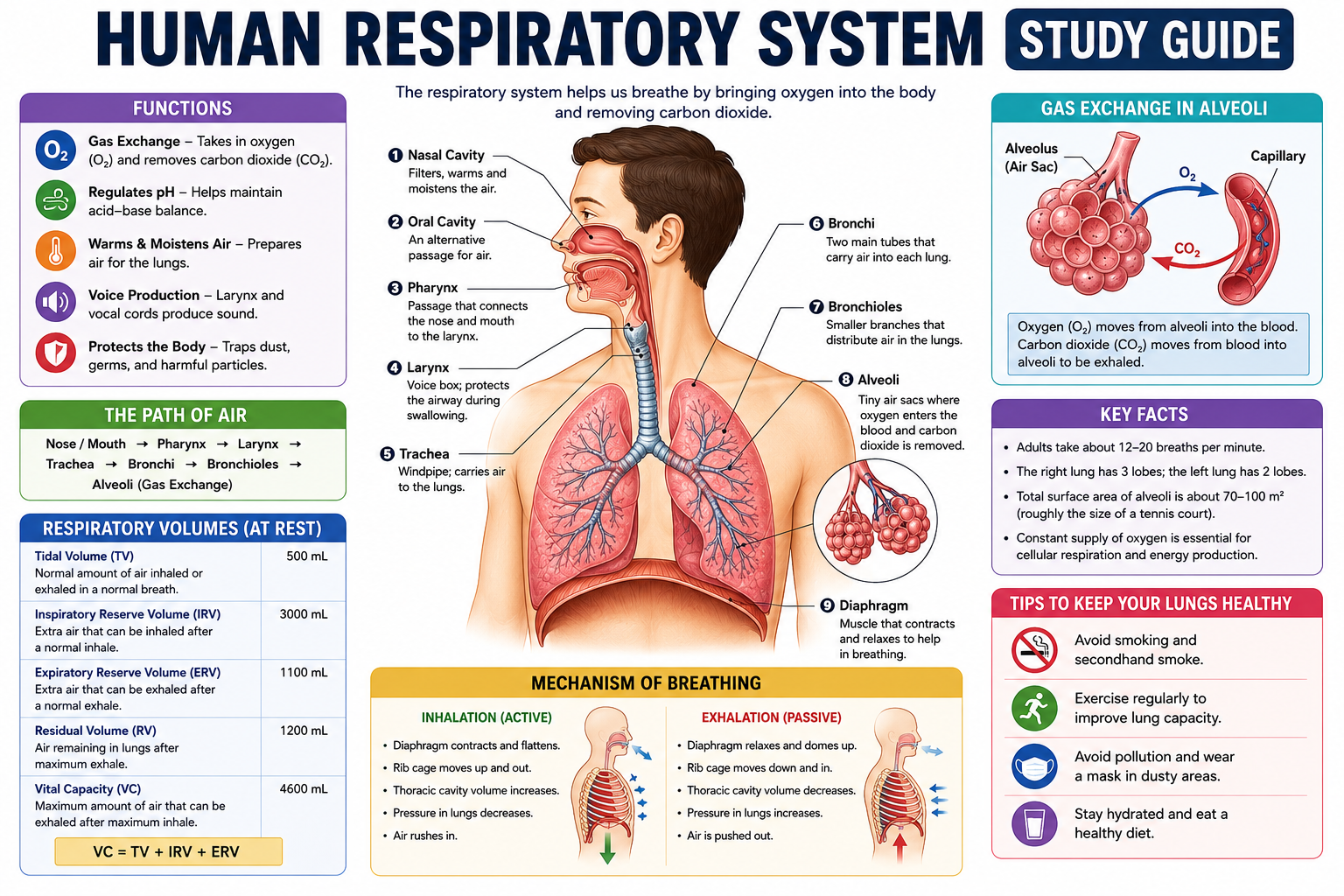
Right now, without any conscious effort on your part, you’re breathing. Your diaphragm is contracting, your lungs are expanding, and somewhere in the depths of your lung tissue, millions of tiny air sacs are exchanging gases with your blood—delivering oxygen to every cell in your body and removing the carbon dioxide those cells produce as metabolic waste. You’ll do this approximately 20,000 times today. And unlike your heartbeat, which you can sometimes hear or feel, breathing happens so smoothly and automatically that you almost never think about it—until something goes wrong.
This human respiratory system study guide is designed to give you a complete, clear, and clinically meaningful understanding of how breathing works, how gas exchange happens, what each respiratory organ does, and what happens when respiratory health fails. Whether you’re preparing for a nursing exam, studying for AP Biology or A-levels, revising for a medical entrance test, or simply trying to understand what’s actually happening when you take a breath, this guide covers everything you need.
The respiratory system is one of the most elegantly designed systems in the human body. It solves a genuinely difficult engineering challenge: how do you continuously supply oxygen—which doesn’t dissolve well in water—to trillions of cells distributed throughout a complex three-dimensional organism, while simultaneously removing the carbon dioxide those cells constantly produce? The answer involves an intricate combination of anatomy, physics, biochemistry, and neural control that we’ll explore step by step.
We’ll cover every respiratory organ, the mechanics of breathing, the physiology of gas exchange, oxygen and carbon dioxide transport in blood, lung volumes and capacities, the brain’s control of breathing, common respiratory disorders, and everything you need for exam success—including practice questions, memory tricks, and a revision checklist.
Take a breath. Let’s begin.
Key Takeaways
By the end of this guide, you’ll be able to:
- Identify and describe the function of every major organ of the respiratory system
- Explain the mechanics of inhalation and exhalation using pressure principles
- Describe gas exchange in the alveoli and explain why it works by diffusion
- Explain how oxygen is transported in blood (primarily as oxyhemoglobin)
- Describe how carbon dioxide is transported (dissolved, carbamino compounds, and bicarbonate)
- Define and distinguish all major lung volumes and capacities
- Explain how the medulla oblongata and pons control breathing rate and depth
- Compare the upper and lower respiratory tracts
- Identify and describe ten common respiratory disorders
- Apply memory tricks and exam strategies for respiratory system assessments
What Is the Human Respiratory System?
The human respiratory system is the organ system responsible for gas exchange—bringing oxygen from the atmosphere into the body and expelling carbon dioxide, the waste product of cellular metabolism. It works in intimate partnership with the circulatory system to ensure that every living cell receives a continuous oxygen supply.
The respiratory system consists of two functional divisions:
1. The conducting zone – A series of passageways that filter, warm, humidify, and transport air from the external environment to the gas exchange surfaces. Includes: nasal cavity, pharynx, larynx, trachea, bronchi, and larger bronchioles. No gas exchange occurs here.
2. The respiratory zone – The actual gas exchange region, consisting of respiratory bronchioles, alveolar ducts, and alveoli—where oxygen moves into the blood and carbon dioxide moves out.
A useful analogy: the conducting zone is like a highway system—it gets the air to where it needs to go, conditioning it along the way. The respiratory zone is like the destination—where the actual work happens.
The respiratory system is also divided anatomically into:
- Upper respiratory tract – Nose, nasal cavity, pharynx, and larynx
- Lower respiratory tract – Trachea, bronchi, bronchioles, and lungs (including alveoli)
Why the Respiratory System Is Important
Every cell in the human body requires a constant supply of oxygen for aerobic cellular respiration—the process by which glucose is converted to ATP (usable energy). Without oxygen, this process fails within seconds in sensitive tissues. Brain cells, for example, begin to die after just 4–6 minutes without oxygen. This is why respiratory failure is a medical emergency and why cardiopulmonary resuscitation (CPR) prioritizes maintaining breathing and circulation simultaneously.
Beyond oxygen delivery, the respiratory system:
- Removes CO₂ – CO₂ is toxic in high concentrations; its accumulation causes respiratory acidosis, which disrupts enzyme function and cellular metabolism throughout the body
- Regulates blood pH – CO₂ dissolves in blood to form carbonic acid; the respiratory system adjusts CO₂ levels to maintain blood pH within the narrow range of 7.35–7.45
- Enables vocalization – Air flowing past the vocal cords in the larynx generates sound; speech and communication depend on respiratory airflow
- Filters and defends – Mucus and cilia trap inhaled particles and pathogens; immune cells in the respiratory tract provide first-line defense against infection
- Maintains body temperature – Exhaled air carries heat and water vapor, contributing to thermoregulation
Main Functions of the Respiratory System
| Function | Description |
|---|---|
| Gas exchange | Deliver O₂ to blood; remove CO₂ from blood at alveoli |
| Ventilation | Move air in and out of the lungs (breathing) |
| Air conditioning | Warm, humidify, and filter incoming air |
| pH regulation | Adjust CO₂ levels to maintain blood pH 7.35–7.45 |
| Vocalization | Generate sound through larynx for speech |
| Olfaction | Enable sense of smell via nasal epithelium |
| Immune defense | Trap pathogens and particles in mucus; clear via cilia |
| Thermoregulation | Exhale heat and water vapor to cool the body |
History of Respiratory System Research
The understanding of breathing and gas exchange has evolved over centuries through remarkable experiments and insights.
Ancient Understanding
Ancient Greek physicians, including Hippocrates and Galen, recognized that air was essential for life but understood its role primarily through the lens of humoral theory. Galen believed the lungs cooled the blood and that “vital spirits” were created in the heart through air contact—a picture that was conceptually wrong but showed early recognition of the lung-heart connection.
Andreas Vesalius (1543)
Vesalius’s anatomical masterwork De Humani Corporis Fabrica provided the first accurate anatomical descriptions of lung structure, correcting many of Galen’s errors. His detailed illustrations of the lungs, trachea, and bronchi remained reference standards for generations.
William Harvey (1628)
Harvey’s demonstration of blood circulation—showing that blood flows from the heart to the lungs and back—established the pulmonary circulation and laid the foundation for understanding how gases could be exchanged between air and blood.
Robert Boyle (1660s)
Boyle’s experiments with air pumps demonstrated that animals die in a vacuum and that air is necessary for combustion—suggesting a connection between breathing and burning (both consume something from air). His work on the relationship between gas pressure and volume (Boyle’s Law) remains essential for understanding breathing mechanics.
Antoine Lavoisier and Joseph Priestley (1770s–1780s)
Priestley discovered oxygen (which he called “dephlogisticated air”) in 1774. Lavoisier named it, showed that it was consumed in breathing and combustion, and that CO₂ was produced—establishing respiration as a form of controlled oxidation. He performed the first measurements of oxygen consumption and CO₂ production during breathing.
20th Century Advances
The discovery of hemoglobin’s oxygen-binding properties, the elucidation of the biochemistry of gas transport, and the development of spirometry (measuring lung volumes) transformed respiratory physiology. Understanding of respiratory control centers in the brainstem emerged through careful neurophysiological experiments in the early-to-mid 20th century.
Structure of the Human Respiratory System
Nose and Nasal Cavity
The nose is the primary entry point for air and the site where incoming air begins its conditioning process. The external nose consists of bone (nasal bones) and cartilage, while the interior—the nasal cavity—is a large space divided by the nasal septum into left and right chambers.
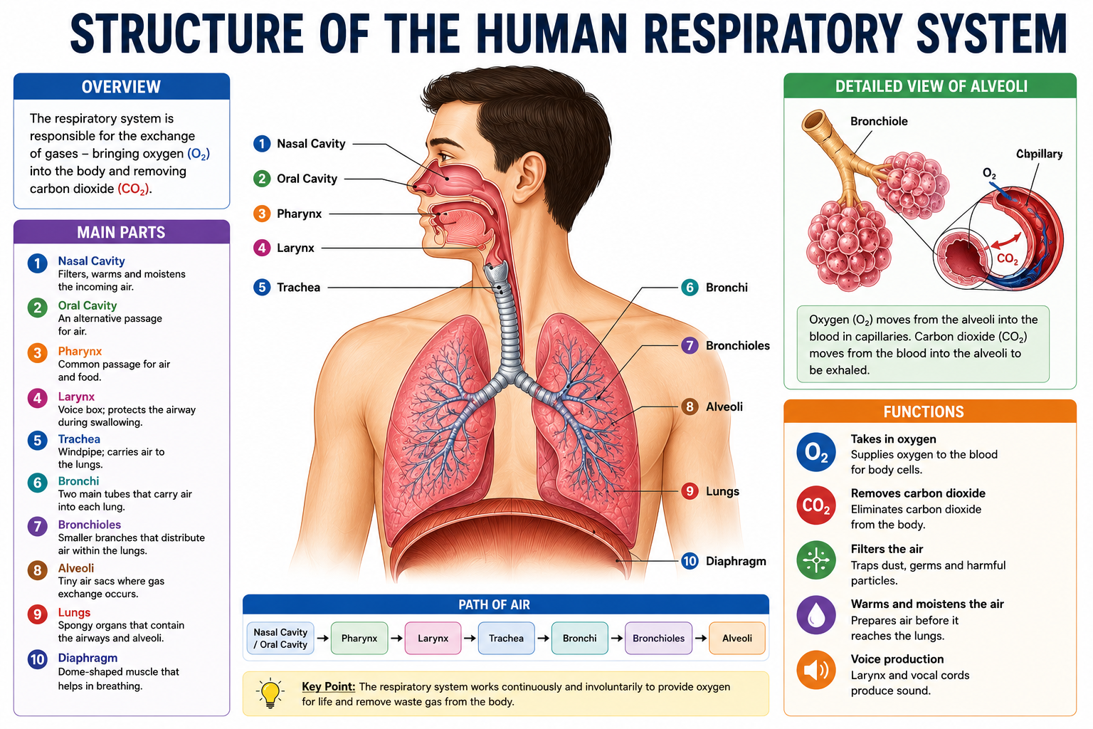
The nasal cavity is lined with:
- Mucous membrane – Secretes mucus that traps dust, pollen, bacteria, and other particles
- Cilia – Hair-like projections that sweep trapped particles toward the throat to be swallowed
- Rich blood supply – Warms incoming cold air to body temperature
- Moist epithelium – Humidifies dry air
Turbinates (nasal conchae) – Three bony shelf-like projections on the lateral walls of the nasal cavity that create turbulence in airflow, maximizing contact between incoming air and the conditioning mucosa. They dramatically increase surface area for air conditioning.
Olfactory epithelium – Located in the upper nasal cavity; contains chemoreceptors for smell that send signals to the brain via the olfactory nerve (cranial nerve I).
The nose also contains nasal hairs (vibrissae) at the entrance—the first mechanical filter for large particles.
Pharynx
The pharynx is a muscular funnel approximately 13 cm long connecting the nasal cavity and mouth to the larynx and esophagus. It serves as a shared passageway for both air and food.
Three regions of the pharynx:
- Nasopharynx – Upper section; air only; houses the pharyngeal tonsil (adenoid) and the openings of the Eustachian tubes (connecting to the middle ear)
- Oropharynx – Middle section; air and food; contains the palatine tonsils
- Laryngopharynx – Lower section; air and food; divides into the larynx (airway) and esophagus (food passage)
Larynx
The larynx (voice box) is a rigid structure made of nine cartilages—the most prominent being the thyroid cartilage (Adam’s apple) and the epiglottis.
Functions of the larynx:
- Vocalization – The vocal cords (vocal folds) span the larynx; air flowing past them causes vibration, producing sound. Muscles adjust tension and length of the cords, controlling pitch.
- Airway protection – The epiglottis acts as a trapdoor: during swallowing, it folds down over the glottis (opening of the larynx), directing food into the esophagus and preventing aspiration into the airway
- Cough reflex – Irritants triggering receptors in the larynx produce a reflexive cough to expel material
Laryngospasm—involuntary spasm of the laryngeal muscles closing the airway—is the mechanism behind “something going down the wrong way,” producing a brief choking sensation.
Trachea
The trachea (windpipe) is a tube approximately 10–12 cm long and 2–2.5 cm in diameter running from the larynx into the chest. It’s reinforced by 16–20 C-shaped rings of hyaline cartilage that prevent the trachea from collapsing during inhalation when pressure inside drops below atmospheric pressure.
The opening of each C-ring faces posteriorly (toward the esophagus), and the posterior wall is composed of smooth muscle and connective tissue (trachealis muscle)—allowing the esophagus to expand into this space when food is swallowed.
The tracheal lining contains pseudostratified ciliated columnar epithelium with goblet cells that produce mucus—continuing the mucociliary clearance system from the nasal cavity. The cilia beat upward (toward the throat) continuously, moving the “mucociliary escalator” of trapped particles out of the lungs to be swallowed or expectorated.
Bronchi
At the level of the 4th–5th thoracic vertebra, the trachea divides at the carina into the left primary bronchus and right primary bronchus—each entering its respective lung.
Key anatomical difference:
- Right primary bronchus – Shorter, wider, and more vertical than the left
- Left primary bronchus – Longer and more horizontal
This is clinically important: inhaled foreign objects (and aspirated vomit, gastric tube misplacements) more commonly enter the right bronchus due to its more vertical angle.
Within the lungs, primary bronchi divide into secondary (lobar) bronchi (3 on the right, 2 on the left—one per lobe), which then divide into tertiary (segmental) bronchi supplying each bronchopulmonary segment. The bronchi contain cartilage in their walls, which maintains patency.
Bronchioles
Bronchioles are the airways that branch from the smallest bronchi. They lack the cartilaginous reinforcement of bronchi—their diameter is controlled entirely by smooth muscle contraction and relaxation.
- Bronchioles – About 1 mm in diameter; controlled by autonomic nervous system. Sympathetic stimulation (epinephrine/norepinephrine) causes bronchodilation (widening); parasympathetic stimulation causes bronchoconstriction (narrowing)
- Terminal bronchioles – The final purely conducting airways; about 0.5 mm in diameter
- Respiratory bronchioles – Have occasional alveoli in their walls; the beginning of the respiratory zone where gas exchange begins
This is why adrenaline (epinephrine) helps during an asthma attack—it triggers sympathetic bronchodilation, widening the airways and improving airflow.
Alveoli
Alveoli are the microscopic, grape-like air sacs at the terminal ends of the respiratory tree. They are the functional units of gas exchange—where oxygen enters the blood and CO₂ leaves.
Key facts about alveoli:
- Approximately 300–500 million alveoli in healthy adult lungs
- Total alveolar surface area: approximately 70–100 m² (about the size of a tennis court)
- Average diameter: ~200–300 micrometers
- Wall thickness: approximately 0.5 micrometers—extremely thin to minimize diffusion distance
Alveolar cell types:
- Type I pneumocytes – Flat, thin cells covering ~95% of the alveolar surface; primary site of gas exchange
- Type II pneumocytes – Cuboidal cells (~5% of surface) that produce surfactant—a phospholipid mixture that reduces surface tension in alveoli, preventing collapse
- Alveolar macrophages – “Dust cells”; phagocytize bacteria, dust, and other inhaled particles that reach the alveoli
Surfactant is critically important. Without it, the alveoli would collapse with each exhalation (because surface tension would cause small spheres to collapse, following the law of Laplace). Premature infants often lack sufficient surfactant, causing Respiratory Distress Syndrome (RDS)—a leading cause of neonatal mortality that is now treated with exogenous surfactant therapy.
Each alveolus is surrounded by a dense network of pulmonary capillaries, and the alveolar-capillary membrane (blood-air barrier) is only about 0.5 micrometers thick—making diffusion of gases extremely rapid.
Lungs
The lungs are a pair of spongy, cone-shaped organs that fill most of the thoracic cavity. Together they weigh approximately 1–1.3 kg.
Lobes:
- Right lung – Three lobes: superior, middle, and inferior; slightly larger than the left lung
- Left lung – Two lobes: superior and inferior; has a concave indentation called the cardiac notch to accommodate the heart
Each lobe is divided into bronchopulmonary segments (10 in the right lung, 8–10 in the left), each with its own segmental bronchus and blood supply—important for surgical resection of diseased segments.
The hilum is the medial surface where the bronchi, pulmonary vessels, lymphatics, and nerves enter and exit the lung.
Pleura
Each lung is surrounded by a double-layered membrane called the pleura:
- Visceral pleura – Directly covers the lung surface
- Parietal pleura – Lines the thoracic wall, diaphragm, and mediastinum
Between the two layers is the pleural cavity, containing a small amount (~10–15 mL) of pleural fluid—a lubricating serous fluid that reduces friction as the lungs expand and contract. The pleural cavity is not a real space under normal conditions—it’s a potential space kept in negative pressure.
Clinical importance of the pleura:
- Pleuritis (pleurisy) – Inflammation of the pleura causing sharp pain with breathing
- Pleural effusion – Accumulation of excess fluid in the pleural cavity, compressing the lung
- Pneumothorax – Air entering the pleural cavity (from lung puncture or chest wound), collapsing the lung
Diaphragm
The diaphragm is the primary muscle of breathing—a dome-shaped sheet of skeletal muscle separating the thoracic and abdominal cavities. It’s innervated by the phrenic nerve (from cervical spinal cord segments C3–C5—remembered by the mnemonic: “C3, 4, 5 keeps the diaphragm alive”).
During inhalation, the diaphragm contracts and flattens downward, increasing thoracic volume and decreasing intrathoracic pressure, drawing air into the lungs.
During exhalation at rest, the diaphragm relaxes and returns to its dome shape, reducing thoracic volume.
The diaphragm has three apertures allowing passage of major structures: the aorta, esophagus, and inferior vena cava.
Intercostal Muscles
The intercostal muscles occupy the spaces between ribs and contribute to breathing, particularly during forceful ventilation:
- External intercostal muscles – Contract during inhalation, elevating the ribs and expanding the chest anterolaterally
- Internal intercostal muscles – Contract during forced exhalation, depressing the ribs and reducing thoracic volume
During normal quiet breathing, only the diaphragm and external intercostals are needed. During exercise or respiratory distress, accessory muscles of respiration activate: sternocleidomastoid and scalenes (elevate the rib cage), and abdominal muscles (force exhalation).
Upper vs Lower Respiratory Tract
| Feature | Upper Respiratory Tract | Lower Respiratory Tract |
|---|---|---|
| Organs included | Nose, nasal cavity, pharynx, larynx | Trachea, bronchi, bronchioles, alveoli, lungs |
| Primary function | Filter, warm, humidify air; vocalization | Gas exchange; air conduction to alveoli |
| Air passage type | Conducting only | Conducting + respiratory (gas exchange) |
| Cartilage | Present in larynx and nose | Trachea, bronchi (absent in bronchioles) |
| Common disorders | Common cold, sinusitis, tonsillitis, laryngitis | Bronchitis, pneumonia, asthma, COPD, tuberculosis |
| Infection frequency | Very common (directly exposed to pathogens) | Less common but more serious |
| Mucus production | Abundant | Present in conducting airways |
| Cilia presence | Throughout | Trachea and bronchi; absent in alveoli |
Journey of Air Through the Respiratory System
EXTERNAL ENVIRONMENT
        ↓
NOSE/MOUTH → Air filtered, warmed, humidified
        ↓
PHARYNX → Common passage for air and food
        ↓
LARYNX → Air-only passage; epiglottis closes during swallowing
        ↓
TRACHEA → Air conducted downward; C-rings maintain patency
        ↓
PRIMARY BRONCHI → Right and left lungs
        ↓
SECONDARY (LOBAR) BRONCHI → Each lung lobe
        ↓
TERTIARY (SEGMENTAL) BRONCHI → Bronchopulmonary segments
        ↓
BRONCHIOLES → Airways <1mm; no cartilage
        ↓
TERMINAL BRONCHIOLES → Final conducting airways
        ↓
RESPIRATORY BRONCHIOLES → Gas exchange begins
        ↓
ALVEOLAR DUCTS
        ↓
ALVEOLI → Primary gas exchange surface
        ↓
O₂ diffuses into pulmonary capillary blood
CO₂ diffuses out of blood into alveolar air
Breathing Process Explained
Breathing (pulmonary ventilation) is the process of moving air into and out of the lungs. It operates entirely on pressure differentials—air always moves from regions of higher pressure to lower pressure.
Inhalation (Inspiration)
- Diaphragm contracts – Moves downward, increasing thoracic cavity volume
- External intercostals contract – Lift ribs up and out, further increasing thoracic volume
- Thoracic volume increases → Intrapulmonary pressure decreases (drops ~1–3 mmHg below atmospheric pressure)
- Pressure gradient created – Atmospheric pressure > intrapulmonary pressure
- Air flows into the lungs down the pressure gradient
This follows Boyle’s Law: when volume increases at constant temperature, pressure decreases. Inhalation is an active process requiring muscular energy.
Exhalation (Expiration)
At rest (passive exhalation):
- Diaphragm relaxes – Returns to dome shape
- External intercostals relax – Ribs drop to resting position
- Thoracic volume decreases → Intrapulmonary pressure increases (rises ~1–3 mmHg above atmospheric pressure)
- Air flows out of the lungs down the new pressure gradient
Quiet exhalation is passive—driven by the elastic recoil of stretched lung tissue, not active muscle contraction.
During forced exhalation (exercise, coughing, playing a wind instrument):
- Internal intercostals and abdominal muscles actively contract
- Dramatically reduces thoracic volume
- Forces air out rapidly and completely
Mechanics of Breathing
Lung compliance – The ease with which lungs and chest wall expand. High compliance = lungs stretch easily (good). Low compliance = stiff lungs requiring more effort to inflate (as in pulmonary fibrosis).
Airway resistance – The opposition to airflow from friction in airways. Higher resistance = more effort required to breathe (as in asthma, where bronchospasm narrows airways).
Elastic recoil – The tendency of stretched lung tissue to spring back to its resting size, driving passive exhalation.
Surfactant’s role in mechanics: Without surfactant, surface tension at the alveolar air-water interface would be enormous, requiring extraordinary effort to inflate alveoli and causing them to collapse on each exhalation. Surfactant reduces surface tension by ~70%, dramatically reducing the work of breathing.
Transmural pressure – The pressure difference between the inside of the alveolus and the pleural space. The negative intrapleural pressure (approximately −4 to −8 mmHg relative to atmospheric) keeps the lungs expanded between breaths.
Gas Exchange in the Alveoli
Gas exchange is driven entirely by diffusion—the passive movement of gases from areas of high partial pressure to areas of low partial pressure. No active transport is required; the physics of diffusion does all the work—provided the diffusion distance is short and the surface area is large. The alveoli satisfy both conditions magnificently.
Partial pressures at the alveolar level:
| Gas | Atmospheric Air | Alveolar Air | Pulmonary Capillary Blood (arriving) | Pulmonary Capillary Blood (leaving) |
|---|---|---|---|---|
| O₂ | 159 mmHg | 100 mmHg | 40 mmHg | 100 mmHg |
| CO₂ | 0.3 mmHg | 40 mmHg | 45 mmHg | 40 mmHg |
Oxygen exchange: pO₂ in alveoli (100 mmHg) > pO₂ in deoxygenated blood arriving at pulmonary capillaries (40 mmHg). Oxygen diffuses from alveoli into blood.
Carbon dioxide exchange: pCO₂ in venous blood arriving at pulmonary capillaries (45 mmHg) > pCO₂ in alveolar air (40 mmHg). CO₂ diffuses from blood into alveoli to be exhaled.
At the tissues (internal respiration): The reverse exchange occurs—pO₂ in arterial blood (100 mmHg) > pO₂ in actively metabolizing tissue cells (as low as 20–40 mmHg in active muscle). Oxygen diffuses from blood into cells. pCO₂ in tissues (45+ mmHg) > pCO₂ in arterial blood (40 mmHg). CO₂ diffuses from cells into blood.
Factors affecting gas exchange efficiency:
- Thickness of alveolar membrane – Thicker = slower diffusion (as in pulmonary fibrosis)
- Surface area – Less surface area = less exchange (as in emphysema, where alveoli are destroyed)
- Partial pressure gradients – Steeper gradient = faster diffusion
- Gas solubility – CO₂ diffuses ~20x faster than O₂ because it’s more soluble in aqueous solutions. This is why CO₂ elimination is not impaired until very late in restrictive lung diseases, long after O₂ uptake becomes deficient.
Oxygen Transport in Blood
Oxygen is transported in blood in two forms:
1. Dissolved in plasma (~1.5%)
A very small amount of O₂ physically dissolves in blood plasma. This is too little to sustain life—it would require blood flow rates 20x the normal cardiac output to meet resting oxygen demands.
2. Bound to hemoglobin (~98.5%)
The vast majority of oxygen is transported by hemoglobin (Hb)—a protein in red blood cells consisting of four subunits, each containing an iron-containing heme group that can bind one O₂ molecule. Each hemoglobin molecule can carry up to 4 oxygen molecules as oxyhemoglobin (HbO₂).
The oxygen-hemoglobin dissociation curve describes the relationship between pO₂ and hemoglobin saturation. It has an S-shaped (sigmoidal) curve with important implications:
- At high pO₂ (like in the lungs, ~100 mmHg): Hemoglobin is nearly fully saturated (~98%)
- At lower pO₂ (like in active tissues, ~40 mmHg): Hemoglobin releases much of its O₂
- The steep portion of the curve ensures large O₂ delivery with modest pO₂ changes in tissues
Factors that shift the curve RIGHT (hemoglobin releases more O₂—beneficial during exercise):
- Increased CO₂ (Bohr effect)
- Decreased pH (acidosis)
- Increased temperature
- Increased 2,3-bisphosphoglycerate (2,3-BPG)
Factors that shift the curve LEFT (hemoglobin holds O₂ more tightly—less delivery to tissues):
- Decreased CO₂
- Increased pH
- Decreased temperature
- Carbon monoxide (CO) poisoning—CO binds hemoglobin ~240x more tightly than O₂, displacing it
Carbon Dioxide Transport
CO₂ is transported from tissues to the lungs in three forms:
1. Dissolved in plasma (~7–10%)
Small amount physically dissolved; contributes to pCO₂ driving diffusion.
2. Carbamino compounds (~20–23%)
CO₂ binds directly to the globin (protein) portions of hemoglobin and plasma proteins, forming carbaminohemoglobin (HbCO₂). This binding is favored in tissues where pCO₂ is high and O₂ saturation is low (deoxygenated hemoglobin carries more CO₂—the Haldane effect).
3. Bicarbonate ions (~70%)
The majority of CO₂ is converted to bicarbonate (HCO₃⁻) inside red blood cells:
CO₂ + H₂O → H₂CO₃ → H⁺ + HCO₃⁻
(catalyzed by carbonic anhydrase)- HCO₃⁻ exits the red blood cell into plasma (in exchange for Cl⁻—the chloride shift)
- H⁺ is buffered by hemoglobin (preventing acidosis)
- At the lungs, the process reverses: HCO₃⁻ re-enters RBCs, combines with H⁺ to reform CO₂, which diffuses into alveolar air
Role of Hemoglobin
Hemoglobin is arguably the most important protein in the respiratory system. Beyond oxygen transport:
- Buffers blood pH – Binds H⁺ ions released during CO₂ hydration, preventing acidosis
- Carries CO₂ as carbaminohemoglobin
- Cooperative binding – Each O₂ binding makes subsequent binding easier (structural change in the molecule); each O₂ release makes subsequent release easier—giving rise to the S-shaped dissociation curve
Hemoglobin variants:
- Fetal hemoglobin (HbF) – Has higher O₂ affinity than adult hemoglobin (HbA), allowing the fetus to extract O₂ from maternal blood across the placenta
- Sickle cell hemoglobin (HbS) – Mutant hemoglobin that polymerizes under low O₂ conditions, causing red blood cells to sickle—distorting shape, blocking capillaries, and reducing O₂ delivery
- Methemoglobin – Iron in heme oxidized from Fe²⁺ to Fe³⁺; cannot bind O₂; certain drugs and chemicals cause methemoglobinemia
Cellular Respiration vs External Respiration
| Feature | External Respiration | Cellular Respiration |
|---|---|---|
| Definition | Gas exchange between lungs and blood (and blood and tissues) | Biochemical process breaking down glucose to produce ATP |
| Location | Lungs (alveoli) and body tissues | Mitochondria of all cells |
| Process type | Physical (diffusion) | Chemical (enzymatic reactions) |
| Oxygen role | Moves from alveoli to blood; blood to cells | Final electron acceptor in ETC |
| CO₂ role | Moves from blood to alveoli for exhalation | Produced as waste product |
| Energy | No ATP produced | ATP produced (~30–32 per glucose) |
| Requires breathing? | Yes | Not directly—uses O₂ delivered by breathing |
| Also called | Pulmonary respiration / gas exchange | Internal respiration (at cellular level) |
Lung Volumes and Lung Capacities
Spirometry—the measurement of lung volumes—is a fundamental clinical and physiological tool. Understanding these values is consistently tested in anatomy, physiology, and nursing exams.
Tidal Volume (TV)
- Definition: Volume of air inhaled or exhaled in one normal, quiet breath
- Normal value: ~500 mL (0.5 L)
- Clinical note: Reduced in restrictive lung diseases
Inspiratory Reserve Volume (IRV)
- Definition: Maximum additional volume of air that can be inhaled AFTER a normal inhalation
- Normal value: ~3,000–3,500 mL (3–3.5 L)
- Used during: Deep breathing, exercise
Expiratory Reserve Volume (ERV)
- Definition: Maximum additional volume of air that can be forcibly exhaled AFTER a normal exhalation
- Normal value: ~1,000–1,200 mL (1–1.2 L)
- Reduced in: Obesity, pregnancy (abdominal contents restrict diaphragm)
Residual Volume (RV)
- Definition: Volume of air remaining in the lungs AFTER maximum forced exhalation
- Normal value: ~1,200–1,500 mL (1.2–1.5 L)
- Cannot be exhaled – Keeps alveoli from collapsing completely; cannot be measured by spirometry alone
- Increased in: Emphysema (air trapping)
Vital Capacity (VC)
- Definition: Maximum volume of air that can be exhaled after maximum inhalation
- Formula: VC = TV + IRV + ERV
- Normal value: ~4,500–5,500 mL (4.5–5.5 L)
- Reduced in: Both restrictive (pulmonary fibrosis) and obstructive (COPD) diseases
Total Lung Capacity (TLC)
- Definition: Total volume of air in the lungs after maximum inhalation
- Formula: TLC = VC + RV
- Normal value: ~6,000 mL (6 L)
Summary Table:
| Volume/Capacity | Abbreviation | Normal Value | Formula |
|---|---|---|---|
| Tidal Volume | TV | ~500 mL | — |
| Inspiratory Reserve Volume | IRV | ~3,000–3,500 mL | — |
| Expiratory Reserve Volume | ERV | ~1,000–1,200 mL | — |
| Residual Volume | RV | ~1,200–1,500 mL | — |
| Inspiratory Capacity | IC | ~3,500–4,000 mL | TV + IRV |
| Functional Residual Capacity | FRC | ~2,200–2,700 mL | ERV + RV |
| Vital Capacity | VC | ~4,500–5,500 mL | TV + IRV + ERV |
| Total Lung Capacity | TLC | ~6,000 mL | VC + RV |
Exam Tip: Residual volume cannot be measured by simple spirometry—it requires body plethysmography or gas dilution techniques. This is a reliable exam question.
Respiratory System Diagram Explained
A complete diagram of the human respiratory system includes:
Upper respiratory tract (labeled top to bottom):
- Nasal cavity (with turbinates)
- Oral cavity
- Pharynx (naso-, oro-, laryngopharynx)
- Epiglottis
- Larynx (with thyroid cartilage and vocal cords)
Lower respiratory tract:
- Trachea (with C-shaped cartilage rings)
- Left and right primary bronchi
- Secondary (lobar) bronchi
- Tertiary (segmental) bronchi
- Bronchioles
- Terminal bronchioles
Lung anatomy:
- Right lung (3 lobes) and Left lung (2 lobes, cardiac notch)
- Pleural layers (visceral and parietal)
- Hilum
Alveolar detail (magnified):
- Alveolus surrounded by pulmonary capillaries
- Alveolar-capillary membrane
- Type I and Type II pneumocytes
- Alveolar macrophages
Breathing mechanism:
- Diaphragm (contracted/flattened during inhalation)
- External intercostal muscles
Human Respiratory System Flowchart
AIR ENTERS → NASAL CAVITY
                    ↓
            Filtered, warmed, humidified
                    ↓
              PHARYNX
                    ↓
              LARYNX (epiglottis protects airway during swallowing)
                    ↓
              TRACHEA
                    ↓
         ┌──────────┴──────────┐
    RIGHT BRONCHUS        LEFT BRONCHUS
         ↓                       ↓
   Secondary bronchi      Secondary bronchi
         ↓                       ↓
   Tertiary bronchi       Tertiary bronchi
         ↓                       ↓
    Bronchioles            Bronchioles
         ↓                       ↓
Terminal bronchioles    Terminal bronchioles
         ↓                       ↓
Respiratory bronchioles  Respiratory bronchioles
         ↓                       ↓
    Alveolar ducts         Alveolar ducts
         ↓                       ↓
      ALVEOLI ←←←←←←←←←←←← ALVEOLI
                    ↓
         GAS EXCHANGE OCCURS
         O₂ → BLOOD
         CO₂ → ALVEOLAR AIR → EXHALED
Respiratory System and Circulatory System Relationship
The respiratory and circulatory systems are so intimately intertwined that they’re often discussed together as a single functional unit—the cardiorespiratory system.
Pulmonary circulation:
- Deoxygenated blood travels from the right ventricle of the heart through the pulmonary artery to the pulmonary capillaries surrounding alveoli
- Gas exchange occurs (O₂ in, CO₂ out)
- Oxygenated blood returns via the pulmonary veins to the left atrium, then left ventricle
- Left ventricle pumps oxygenated blood to the body (systemic circulation)
Systemic circulation:
- Oxygenated blood travels from left ventricle through the aorta to all body tissues
- At tissue capillaries: O₂ diffuses into cells; CO₂ diffuses into blood
- Deoxygenated blood returns via veins to the right atrium → right ventricle → pulmonary circulation
Ventilation-perfusion (V/Q) matching: For efficient gas exchange, alveolar ventilation (airflow) and pulmonary blood flow (perfusion) must be matched. In lung disease, mismatching can occur—ventilated alveoli not perfused with blood, or perfused capillaries next to non-ventilated alveoli—causing hypoxemia (low blood oxygen) despite breathing.
Respiratory Control by the Brain
Breathing is unusual among body functions—it can be both automatic and voluntary. You can consciously hold your breath or breathe faster, but the moment you lose consciousness (or stop trying to control it), automatic breathing resumes. This dual control is mediated by overlapping brain centers.
Medulla Oblongata
The medulla oblongata contains the primary respiratory control centers:
- Dorsal respiratory group (DRG) – Sets the baseline breathing rhythm; generates the respiratory pattern during quiet breathing; sends inspiratory signals to the diaphragm and external intercostals
- Ventral respiratory group (VRG) – Inactive during quiet breathing; activated during forced ventilation and exercise to recruit accessory muscles of inspiration and expiration
Central chemoreceptors in the medulla detect changes in CO₂ and pH in cerebrospinal fluid (CSF). Rising CO₂ → increased H⁺ in CSF → stimulates medullary chemoreceptors → increased breathing rate and depth to blow off excess CO₂. This is the primary driver of breathing rate—CO₂ levels, not O₂.
Pons
The pons contains centers that fine-tune breathing rhythm:
- Pneumotaxic center (pontine respiratory group) – Sends inhibitory signals to the DRG, limiting inhalation duration and regulating breathing rate; prevents the lungs from being overfilled
- Apneustic center – Sends stimulatory signals to prolong inhalation; counterbalanced by the pneumotaxic center
Peripheral chemoreceptors:
- Carotid bodies – Located at the bifurcation of the common carotid arteries; sensitive primarily to hypoxia (low pO₂), but also to increased pCO₂ and decreased pH
- Aortic bodies – Located in the aortic arch; similar function
Unlike CO₂, O₂ levels must fall substantially (pO₂ < 60 mmHg) before peripheral chemoreceptors significantly stimulate breathing. In patients with chronic hypercapnia (chronically elevated CO₂—as in severe COPD), the respiratory drive becomes increasingly driven by hypoxic stimulation from peripheral chemoreceptors—the hypoxic drive. This is the basis of the concern about giving high-flow oxygen to COPD patients (potentially reducing their breathing drive).
Common Respiratory Disorders
Asthma
Cause: Chronic inflammatory airway disease with bronchial hyperresponsiveness; triggers include allergens (pollen, dust mites), exercise, cold air, infections, and irritants.
Pathophysiology: Inflammation → bronchospasm (bronchoconstriction) + mucus hypersecretion → airway narrowing → increased airway resistance → wheeze, dyspnea.
Symptoms: Episodic wheeze, shortness of breath, chest tightness, cough (especially at night/early morning).
Treatment: Short-acting β₂ agonists (SABA, e.g., salbutamol/albuterol) for acute relief; inhaled corticosteroids (ICS) for long-term control; avoidance of triggers.
Bronchitis
Acute bronchitis: Usually viral; inflammation of bronchial mucosa; productive cough, low-grade fever; self-limiting.
Chronic bronchitis: Defined as productive cough for ≥3 months per year for ≥2 consecutive years; caused by long-term smoking or pollution; chronic mucus hypersecretion; “blue bloater” phenotype in COPD.
Pneumonia
Cause: Infection of lung parenchyma by bacteria (Streptococcus pneumoniae most common), viruses, or fungi; causes alveoli to fill with fluid and inflammatory cells.
Symptoms: Fever, productive cough, dyspnea, pleuritic chest pain, crackles on auscultation.
Treatment: Antibiotics (for bacterial pneumonia); rest, hydration; severe cases require hospitalization and supplemental O₂.
Tuberculosis (TB)
Cause: Mycobacterium tuberculosis; transmitted by respiratory droplets; infects lung tissue, causing granuloma formation.
Symptoms: Chronic productive cough (often blood-tinged), night sweats, weight loss, fever.
Treatment: Long-term multi-drug antibiotic regimen (typically 6 months); drug-resistant TB (MDR-TB) is a major global health challenge.
Influenza
Cause: Influenza A or B viruses; highly contagious; transmitted by respiratory droplets.
Symptoms: Sudden fever, myalgia, headache, cough, fatigue; typically more severe than common cold.
Treatment: Antiviral medications (oseltamivir) if given early; otherwise supportive; annual vaccination is the primary preventive measure.
COVID-19
Cause: SARS-CoV-2 coronavirus; uses ACE2 receptors expressed abundantly in lung epithelium.
Respiratory manifestations: Range from mild upper respiratory symptoms to severe pneumonia, ARDS (Acute Respiratory Distress Syndrome), and respiratory failure requiring mechanical ventilation.
Long COVID: Persistent dyspnea, fatigue, and cognitive symptoms months after acute infection; mechanism involves ongoing inflammation and microvasculature damage.
COPD (Chronic Obstructive Pulmonary Disease)
Cause: Long-term smoking (primary), occupational dust and chemical exposure, air pollution; characterized by chronic airflow obstruction that is not fully reversible.
Two main phenotypes:
- Chronic bronchitis – Mucus hypersecretion, productive cough, “blue bloater”
- Emphysema – Alveolar destruction, air trapping, “pink puffer”
Diagnosis: Spirometry shows FEV₁/FVC ratio < 0.7 (reduced ability to exhale quickly).
Treatment: Smoking cessation (most important), bronchodilators, ICS, pulmonary rehabilitation, supplemental O₂ in severe cases.
Lung Cancer
Cause: Primarily tobacco smoking (~85% of cases); also radon gas, asbestos, air pollution; two main types: non-small cell lung cancer (NSCLC—~85%) and small cell lung cancer (SCLC—~15%).
Symptoms: Often asymptomatic until advanced; persistent cough, hemoptysis, weight loss, chest pain, dyspnea.
Treatment: Surgery, chemotherapy, radiation, targeted therapies (for specific mutations), immunotherapy.
Emphysema
Cause: Permanent destruction of alveolar walls due to chronic inflammation (primarily from smoking); the enzyme elastase from inflammatory cells destroys the elastic tissue in alveolar walls; normally counterbalanced by α₁-antitrypsin (deficiency is a genetic cause of early emphysema).
Effect: Loss of alveolar surface area (reduced gas exchange) and loss of elastic recoil (air trapping, barrel chest).
Symptoms: Progressive dyspnea, reduced exercise tolerance, “pink puffer” appearance.
Pulmonary Fibrosis
Cause: Progressive scarring (fibrosis) of lung tissue, replacing functional alveolar tissue with stiff, non-compliant scar tissue; can be idiopathic (IPF) or secondary (autoimmune diseases, drug reactions, exposure to asbestos or silica).
Effect: Restrictive lung disease—reduced lung compliance, reduced all lung volumes, impaired gas exchange.
Symptoms: Progressive dyspnea, dry cough, “Velcro” crackles on auscultation.
Treatment: Limited; antifibrotic drugs slow progression; lung transplantation in eligible patients.
Respiratory Health Tips
- Don’t smoke—and if you do, get support to quit. Smoking is responsible for ~85% of lung cancers and is the primary cause of COPD. Even secondhand smoke causes significant respiratory harm.
- Avoid air pollutants—check air quality indexes; wear appropriate masks in polluted or occupational environments with dust or chemicals.
- Exercise regularly—aerobic exercise improves lung efficiency, increases respiratory muscle strength, and improves the ventilation-perfusion matching.
- Stay vaccinated—influenza and pneumococcal vaccines significantly reduce the risk of severe respiratory infections.
- Practice nasal breathing where possible—the nose conditions air far more effectively than mouth breathing.
- Maintain indoor air quality—use ventilation, avoid indoor smoking, control humidity (excess moisture promotes mold), use air filters if needed.
- Manage allergies and asthma proactively—don’t wait until symptoms are severe; follow medication plans consistently.
- Practice diaphragmatic breathing—especially beneficial for those with anxiety or chronic respiratory conditions; improves breathing efficiency.
Foods That Support Lung Health
- Omega-3 rich foods (fatty fish, flaxseed, walnuts) – Anti-inflammatory; evidence suggests reduced risk of COPD and improved asthma outcomes
- Fruits rich in antioxidants (berries, citrus, apples) – Quercetin and vitamin C support lung function; apple consumption associated with better lung function in population studies
- Cruciferous vegetables (broccoli, Brussels sprouts) – Contain sulforaphane; anti-inflammatory and anti-carcinogenic properties
- Ginger and turmeric – Anti-inflammatory compounds that may reduce airway inflammation
- Garlic – Contains allicin; antimicrobial and anti-inflammatory properties
- Green tea – Rich in catechins with antioxidant and anti-inflammatory effects
Harmful Habits That Damage the Lungs
- Smoking and vaping – Most damaging; tobacco smoke contains over 7,000 chemicals including carcinogens, causes chronic inflammation and oxidative damage to airways and alveoli
- Burning wood or biomass indoors – Major cause of chronic lung disease globally, particularly in low-income settings
- Occupational exposures – Asbestos (mesothelioma, pulmonary fibrosis), silica (silicosis), coal dust (pneumoconiosis)
- Sedentary lifestyle – Reduces respiratory muscle strength and efficiency
- Chronic mouth breathing – Bypasses nasal conditioning; may increase airway inflammation
Respiratory System in Children
Children’s respiratory systems differ significantly from adults, with important clinical implications:
- Smaller airways – Children have proportionally narrower airways; swelling from infection or inflammation causes proportionally greater airway resistance than in adults
- Higher respiratory rate – Newborns breathe ~40–60 times/minute; adults breathe 12–20 times/minute; rate decreases with age as lung capacity grows relative to oxygen demand
- Horizontal ribs – Infants have more horizontal ribs (vs adults’ downward-sloping ribs), making them more dependent on diaphragmatic breathing; abdominal distension (e.g., from swallowed air) can significantly impair breathing
- Absent surfactant deficiency in preterm infants – Surfactant production begins around 28–32 weeks gestation; premature infants before this gestational age are at risk for Respiratory Distress Syndrome
- Common infections – Viral lower respiratory infections (bronchiolitis from RSV, croup) are leading causes of childhood hospitalization
Respiratory Changes During Aging
The aging respiratory system undergoes predictable changes that reduce reserve capacity:
- Reduced chest wall compliance – Calcification of costal cartilages makes chest wall stiffer; more work required for breathing
- Reduced lung elasticity – Loss of elastic tissue (similar to mild emphysema); air trapping increases; residual volume increases
- Weakened respiratory muscles – Reduced diaphragm and intercostal muscle strength; reduced maximum ventilatory capacity
- Loss of alveolar surface area – Some alveolar coalescence with aging; reduced maximum gas exchange capacity
- Reduced chemoreceptor sensitivity – Blunted responses to hypoxia and hypercapnia; reduced ability to compensate for respiratory challenges
- Reduced mucociliary clearance – Cilia become less effective; mucus clearance slows; increased vulnerability to pneumonia (a leading cause of death in the elderly)
- All lung volumes change – TLC relatively preserved; FRC and RV increase; vital capacity decreases
Interesting Facts About the Respiratory System
Important Facts Box:
- You breathe approximately 20,000 times per day at rest
- Your lungs contain approximately 300–500 million alveoli—providing a gas exchange surface of ~70–100 m² (the size of a tennis court)
- Each lung contains approximately 2,400 km (1,500 miles) of airways if stretched end to end
- The left lung is approximately 10% smaller than the right to accommodate the heart
- Air stays in the alveoli for less than one second during normal breathing—yet gas exchange is essentially complete in that time
- CO₂ diffuses across the alveolar membrane ~20x faster than O₂ due to its greater solubility
- The mucociliary escalator moves mucus at approximately 1–2 cm per minute—clearing the entire tracheal surface in about an hour
- A healthy adult can exhale at speeds up to 160 km/h during a cough
- Your nose can detect over 1 trillion different scents
- The phrenic nerve, which controls the diaphragm, originates from cervical spinal cord levels C3–C5—which is why high cervical spinal cord injuries (above C4) cause respiratory failure
Common Biology Terms Every Student Should Know
| Term | Definition |
|---|---|
| Ventilation | Movement of air into and out of the lungs (breathing) |
| Respiration | In physiology: gas exchange; in biochemistry: ATP production from glucose |
| Alveolus | Microscopic air sac in the lung; primary gas exchange unit |
| Surfactant | Phospholipid produced by Type II pneumocytes; reduces alveolar surface tension |
| Pleura | Double-layer membrane surrounding each lung |
| Pneumothorax | Air in the pleural cavity causing lung collapse |
| Intrapleural pressure | Pressure in the pleural cavity; normally negative (~−4 to −8 mmHg) |
| Tidal volume | Volume of air in one normal breath (~500 mL) |
| Vital capacity | Maximum air exhaled after maximum inhalation |
| Partial pressure | Pressure exerted by one gas in a mixture of gases |
| Oxyhemoglobin | Hemoglobin with bound oxygen |
| Carbaminohemoglobin | Hemoglobin with bound CO₂ |
| Bohr effect | CO₂/H⁺ reduces hemoglobin’s O₂ affinity, promoting O₂ release in tissues |
| Haldane effect | Deoxygenated hemoglobin carries more CO₂ than oxygenated hemoglobin |
| Spirometry | Measurement of lung volumes and capacities |
| FEV₁ | Forced expiratory volume in 1 second; marker of airway obstruction |
| V/Q ratio | Ventilation-perfusion ratio; measure of gas exchange efficiency |
| Hypoxia | Inadequate oxygen delivery to tissues |
| Hypercapnia | Elevated blood CO₂ |
| Cyanosis | Bluish discoloration of skin/mucous membranes due to hypoxemia |
Common Mistakes Students Make
1. Confusing respiration with breathing
“Respiration” has two different meanings depending on context—always specify which you mean. Pulmonary ventilation (breathing) is the physical movement of air. Cellular respiration is the biochemical production of ATP. External respiration is gas exchange at the lungs. These are three different things and confusing them is the most common source of errors in respiratory physiology questions.
2. Saying oxygen drives breathing rate
The primary driver of breathing rate is CO₂—specifically, rising CO₂ detected by central chemoreceptors in the medulla. Oxygen levels must fall substantially (pO₂ < 60 mmHg) before peripheral chemoreceptors significantly stimulate breathing. This is a reliable trick question on exams.
3. Confusing the right and left bronchi differences
The right primary bronchus is shorter, wider, and more vertical. The left is longer, narrower, and more horizontal. The clinical implication: inhaled foreign objects more commonly lodge in the right bronchus. Get these backwards and you’ll lose marks.
4. Saying exhalation is always active
Quiet/normal exhalation is passive—it requires no muscle contraction; it’s driven by elastic recoil of the lung tissue. Only forced exhalation (during exercise, coughing, playing instruments) requires active muscular effort (internal intercostals, abdominals). Students often incorrectly state that exhalation always requires muscular effort.
5. Stating that residual volume can be measured by spirometry
Residual volume cannot be measured by spirometry alone—you can’t exhale it, so a spirometer can’t measure it. It requires body plethysmography or gas dilution techniques. This distinction is tested in virtually every anatomy and physiology assessment that covers lung volumes.
6. Saying the left lung has three lobes
The left lung has two lobes (superior and inferior). The right has three (superior, middle, inferior). Remember: Left = 2 (like the two-chambered right side of the body position it accommodates) or think “Left lungs Lost a lobe to make room for the heart.”
Memory Tricks to Remember Respiratory Organs
Order of air passage (mouth to alveoli):
“Nasty Pharaohs Like Tummy Bugs, But Babies Are Lovely”
→ Nasal cavity, Pharynx, Larynx, Trachea, Bronchi, Bronchioles, Alveolar ducts, Alveoli, Lungs
Lung lobes:
- Right = 3 lobes (3 letters in RIGHT)
- Left = 2 lobes (2 letters in… actually use: Left has Less lobes
Phrenic nerve origin: “C3, 4, 5 keeps the diaphragm alive”
Lung volumes in order (smallest to largest):
“Tiny Inexperienced Explorers Rarely Visit Total Capacity”
→ Tidal volume, IRV, ERV, Residual volume, Vital Capacity, Total lung Capacity
CO₂ transport forms (smallest to largest percentage):
“Dissolved Carries Best” → Dissolved (7–10%), Carbamiho (20–23%), Bicarbonate (70%)
Bohr effect (what increases O₂ release from Hb):
CADET stands at ATTENTION:
→ CO₂ ↑, Acid (pH ↓), DPG (2,3-BPG) ↑, Exercise (temperature ↑) = right shift = more O₂ release
Exam Tips
Exam Strategy Box:
- Know the anatomy in order—from nose to alveoli; be able to list every structure air passes through in sequence
- For gas exchange questions: Always think about partial pressure gradients—O₂ moves from high (alveoli, 100 mmHg) to low (venous blood, 40 mmHg); CO₂ moves from high (venous blood, 45 mmHg) to low (alveoli, 40 mmHg)
- For lung volume questions: Know the formula for vital capacity (TV + IRV + ERV) and total lung capacity (VC + RV); remember RV cannot be measured by spirometry
- For breathing mechanics: Know Boyle’s Law; remember inhalation is active (muscle contraction increases volume → decreases pressure → air flows in) and quiet exhalation is passive
- For disorder questions: Know the mechanism—not just the name and symptoms—asthma = bronchoconstriction; emphysema = alveolar destruction; fibrosis = restrictive disease
- For transport questions: O₂ is mostly as oxyhemoglobin (98.5%); CO₂ is mostly as bicarbonate (70%)
- Draw diagrams under exam conditions—practice drawing the respiratory system from memory; diagrams earn marks and demonstrate understanding
Human Respiratory System Practice Questions
20 Multiple Choice Questions with Answers
- Which structure prevents food from entering the airway during swallowing?
- A) Uvula
- B) Soft palate
- C) Epiglottis ✓
- D) Vocal cords
- The primary driver of breathing rate is:
- A) Oxygen levels in blood
- B) Nitrogen levels
- C) Carbon dioxide levels ✓
- D) pH of urine
- Which cells produce surfactant in the alveoli?
- A) Type I pneumocytes
- B) Type II pneumocytes ✓
- C) Alveolar macrophages
- D) Clara cells
- The right primary bronchus is different from the left because it is:
- A) Longer and narrower
- B) Shorter, wider, and more vertical ✓
- C) Connected to two lung lobes
- D) Lacking cartilage
- Residual volume is defined as:
- A) Volume of air in a normal breath
- B) Maximum air exhaled after maximum inhalation
- C) Air remaining after maximum exhalation ✓
- D) Total volume at maximum inhalation
- The Bohr effect describes:
- A) CO₂ production in red blood cells
- B) Reduced hemoglobin-O₂ affinity in the presence of CO₂ and H⁺ ✓
- C) Diffusion of O₂ across the alveolar membrane
- D) Increased breathing rate in response to hypoxia
- Where do the primary bronchi divide from the trachea?
- A) At the level of the thyroid cartilage
- B) At the carina ✓
- C) At the level of the 1st thoracic vertebra
- D) At the cricoid cartilage
- Which law explains why lung volume increases during inhalation causes air pressure inside to decrease?
- A) Henry’s Law
- B) Dalton’s Law
- C) Fick’s Law
- D) Boyle’s Law ✓
- The majority of CO₂ is transported in blood as:
- A) Dissolved CO₂
- B) Carbaminohemoglobin
- C) Bicarbonate ions ✓
- D) Carbonic acid
- Normal tidal volume at rest is approximately:
- A) 250 mL
- B) 500 mL ✓
- C) 1,000 mL
- D) 1,500 mL
- Which brain region sets the basic rhythmic pattern of breathing?
- A) Cerebellum
- B) Cerebral cortex
- C) Medulla oblongata ✓
- D) Thalamus
- How many lobes does the right lung have?
- A) 2
- B) 3 ✓
- C) 4
- D) 5
- Normal quiet exhalation is:
- A) Active—requires internal intercostal contraction
- B) Passive—driven by elastic recoil ✓
- C) Active—requires abdominal muscle contraction
- D) Passive—driven by diaphragm relaxation and active muscle contraction equally
- Emphysema causes respiratory problems primarily by:
- A) Narrowing the airways through bronchoconstriction
- B) Filling alveoli with fluid
- C) Destroying alveolar walls, reducing surface area ✓
- D) Causing airway inflammation and mucus hypersecretion
- The phrenic nerve innervates which primary breathing muscle?
- A) External intercostals
- B) Internal intercostals
- C) Diaphragm ✓
- D) Sternocleidomastoid
- Vital capacity equals:
- A) Tidal volume + Residual volume
- B) Tidal volume + IRV + ERV ✓
- C) Total lung capacity + Residual volume
- D) IRV + ERV only
- Which structure is described as the “voice box”?
- A) Trachea
- B) Pharynx
- C) Larynx ✓
- D) Bronchus
- Gas exchange in the alveoli is driven by:
- A) Active transport requiring ATP
- B) Osmosis
- C) Pressure gradients causing diffusion ✓
- D) Carrier proteins in the alveolar membrane
- In COPD, the spirometry result characteristically shows:
- A) Normal FEV₁/FVC ratio
- B) FEV₁/FVC ratio < 0.7 ✓
- C) Increased vital capacity
- D) Normal total lung capacity
- Carbon monoxide poisoning is dangerous because CO:
- A) Destroys alveoli
- B) Blocks carbonic anhydrase
- C) Binds hemoglobin much more tightly than O₂, displacing it ✓
- D) Causes bronchoconstriction
10 Short Answer Questions
- Describe the three ways CO₂ is transported in the blood from tissues to the lungs. State the approximate percentage of each form.
- Explain why quiet exhalation is passive but forced exhalation is active. Describe the muscles involved in each.
- Using partial pressures, explain how O₂ moves from alveoli to blood, and how CO₂ moves from blood to alveoli. Why does this process not require active transport?
- Explain the role of surfactant in maintaining alveolar stability. What happens in premature infants who lack sufficient surfactant?
- Describe the oxygen-hemoglobin dissociation curve. What does its S-shape signify, and what shifts it to the right? What is the clinical significance of a rightward shift?
- Explain the difference between the upper and lower respiratory tracts. Which disorders most commonly affect each region?
- Describe the neural control of breathing rate. Which chemoreceptors are most important for regulating normal breathing, and what do they detect?
- A patient has emphysema. Explain how alveolar destruction affects: (a) gas exchange, (b) elastic recoil, (c) residual volume, and (d) the clinical appearance of the patient.
- Define and give normal values for: tidal volume, vital capacity, and total lung capacity. Explain why residual volume cannot be measured by spirometry.
- Compare asthma and COPD in terms of underlying pathophysiology, reversibility of airflow obstruction, typical age of onset, and primary treatment approach.
5 Long Answer Questions
- Describe the complete journey of an oxygen molecule from the atmosphere to a mitochondrion in a working muscle cell. Include every structure it passes through, the mechanism by which it moves at each step (diffusion, bulk flow, etc.), and the partial pressures involved at key locations. Explain the role of hemoglobin in this journey and describe what happens to the hemoglobin once it releases O₂ at the tissues.
- Describe the mechanics of breathing in detail. Explain the role of Boyle’s Law, the changes in lung volume and intrapulmonary pressure during inhalation and exhalation, the muscles involved in each phase, and the difference between quiet and forced breathing. Explain the concepts of lung compliance, airway resistance, and elastic recoil and how disease processes like asthma and pulmonary fibrosis alter these properties.
- Describe the structure of the alveolus in complete detail. Include all cell types and their functions, the alveolar-capillary membrane, the role of surfactant, and the factors that determine the rate of gas exchange (reference Fick’s Law of diffusion). Explain how emphysema and pulmonary fibrosis each differently affect the efficiency of gas exchange and why CO₂ is less affected than O₂ in early restrictive lung disease.
- Describe the neural and chemical control of breathing. Explain the roles of the medulla oblongata and pons. Describe both central and peripheral chemoreceptors—what they detect, where they are located, and how they modify breathing. Explain the clinical significance of the hypoxic drive in COPD patients and why giving these patients high-flow oxygen requires careful consideration.
- Choose five common respiratory disorders and discuss each comprehensively. For each condition, describe: the causative mechanism or etiology, the specific anatomical structures affected, the pathophysiological process causing symptoms, the main clinical features, and the primary treatment approaches. Where relevant, compare related conditions (e.g., asthma vs COPD; emphysema vs chronic bronchitis).
Revision Notes
Quick Revision Summary:
Air pathway: Nose → Pharynx → Larynx → Trachea → Primary bronchi → Secondary bronchi → Tertiary bronchi → Bronchioles → Terminal bronchioles → Respiratory bronchioles → Alveolar ducts → Alveoli
Breathing mechanics: Boyle’s Law; Inhalation = diaphragm contracts (flattens) + external intercostals contract → volume ↑ → pressure ↓ → air flows in (ACTIVE). Quiet exhalation = passive (elastic recoil).
Gas exchange: Driven by partial pressure gradients; O₂ moves alveoli → blood (100→40 mmHg); CO₂ moves blood → alveoli (45→40 mmHg).
O₂ transport: 1.5% dissolved; 98.5% as oxyhemoglobin.
CO₂ transport: 7–10% dissolved; 20–23% carbaminohemoglobin; 70% bicarbonate.
Lung volumes: TV=500mL; VC=TV+IRV+ERV (~4.5–5.5L); TLC=VC+RV (~6L); RV cannot be measured by spirometry.
Breathing control: Primary driver = CO₂ (central chemoreceptors in medulla); O₂ (peripheral chemoreceptors—carotid/aortic bodies) only when severely low.
Lung lobes: Right = 3 (superior, middle, inferior); Left = 2 (superior, inferior—cardiac notch).
Revision Checklist
Work through this honestly before any exam on the respiratory system.
- I can name all structures air passes through from nose to alveoli in correct order
- I can describe the function of each respiratory structure
- I can compare the upper and lower respiratory tracts across key features
- I can explain the mechanics of inhalation and exhalation using Boyle’s Law
- I can distinguish active from passive processes in breathing
- I can explain gas exchange at the alveoli using partial pressure values
- I can describe O₂ transport in blood (two forms with percentages)
- I can describe CO₂ transport in blood (three forms with percentages)
- I can explain the Bohr effect and Haldane effect
- I can define all lung volumes and capacities with normal values
- I can explain why residual volume cannot be measured by spirometry
- I can describe the neural control of breathing (medulla and pons)
- I can explain what drives normal breathing rate (CO₂, not O₂)
- I can describe ten common respiratory disorders (cause, mechanism, symptoms, treatment)
- I have completed at least 20 MCQs and 3 long answer questions from this guide
Best Books for Learning Human Anatomy
- “Anatomy & Physiology” by OpenStax (free online) – Comprehensive, peer-reviewed, and freely accessible. The respiratory system chapters are well-illustrated and cover anatomy, physiology, and clinical applications at an appropriate level for nursing and health science students.
- “Human Anatomy & Physiology” by Marieb and Hoehn – The gold standard undergraduate anatomy and physiology textbook. Used in nursing programs and pre-medical courses worldwide; exceptional respiratory system coverage with outstanding clinical correlation.
- “Guyton and Hall Textbook of Medical Physiology” – The authoritative medical physiology reference. The pulmonary physiology section—particularly the chapters on gas exchange, ventilation, and respiratory control—are among the clearest in medical education literature.
- “West’s Respiratory Physiology: The Essentials” by West and Luks – The definitive concise reference for respiratory physiology, used in medical schools globally. Exceptional for understanding gas exchange, V/Q relationships, and clinical applications. Every serious respiratory physiology student should have a copy.
- “Gray’s Anatomy for Students” – Outstanding anatomical detail with clinical notes and beautiful illustrations. For understanding the three-dimensional relationships of the respiratory structures within the thorax, nothing is clearer.
Free Online Anatomy Resources
- OpenStax Anatomy & Physiology – Respiratory System – Free, peer-reviewed comprehensive coverage of all respiratory anatomy and physiology topics with review questions.
- Khan Academy – Respiratory System – Excellent video lessons covering breathing mechanics, gas exchange, hemoglobin, and lung diseases. Outstanding for visual learners.
- Biology LibreTexts – Respiratory System – Open-access academic content covering respiratory anatomy and physiology at introductory through intermediate levels.
- MedlinePlus – Lung Diseases – US National Library of Medicine’s authoritative resource for respiratory disorders. Clear explanations of all major lung diseases with links to research.
- NCBI Bookshelf – Respiratory Physiology – Free access to authoritative respiratory physiology content. Appropriate for advanced students and medical candidates requiring depth beyond introductory texts.
Frequently Asked Questions
1. What is the primary function of the respiratory system?
The primary function is gas exchange—delivering oxygen from the atmosphere to the blood and removing carbon dioxide (the waste product of cellular metabolism) from the blood to the atmosphere. This is accomplished through the continuous process of ventilation (breathing) and diffusion at the alveoli.
2. What structures make up the upper respiratory tract?
The upper respiratory tract includes the nose, nasal cavity, pharynx (nasopharynx, oropharynx, laryngopharynx), and larynx. These structures filter, warm, and humidify incoming air and serve as the passageway to the lower respiratory tract.
3. What is the role of surfactant?
Surfactant is a phospholipid mixture produced by Type II pneumocytes in the alveoli. It reduces surface tension at the air-liquid interface inside alveoli, preventing them from collapsing during exhalation. Without surfactant, the work of breathing would be enormous and alveoli would collapse. Premature infants with insufficient surfactant develop Respiratory Distress Syndrome.
4. Why does breathing rate increase during exercise?
During exercise, muscles produce more CO₂. Rising blood CO₂ is detected by central chemoreceptors in the medulla oblongata, which increase the frequency and depth of breathing to expel excess CO₂ and bring in more O₂. Lactic acid produced during intense exercise also lowers blood pH, further stimulating breathing.
5. What is the difference between external and internal respiration?
External respiration (pulmonary respiration) is gas exchange between the alveoli and pulmonary capillaries—O₂ moves into blood, CO₂ moves out, at the lungs. Internal respiration is gas exchange between systemic capillaries and body tissues—O₂ moves from blood into cells, CO₂ moves from cells into blood.
6. How is oxygen transported in the blood?
Approximately 98.5% of oxygen is transported bound to hemoglobin as oxyhemoglobin (HbO₂) inside red blood cells. About 1.5% is dissolved directly in plasma. Hemoglobin is essential—without it, blood oxygen-carrying capacity would be far too low to sustain aerobic metabolism.
7. What causes asthma?
Asthma is a chronic inflammatory airway disease characterized by bronchial hyperresponsiveness. Exposure to triggers (allergens, exercise, cold air, viral infections, irritants) causes inflammation, bronchoconstriction (smooth muscle spasm), and mucus hypersecretion—narrowing airways and increasing resistance. The narrowing causes the characteristic wheeze.
8. How does emphysema differ from chronic bronchitis?
Both are components of COPD. Emphysema involves destruction of alveolar walls (loss of surface area for gas exchange and loss of elastic recoil—causing air trapping). Chronic bronchitis involves inflammation and mucus hypersecretion in the bronchi (not alveolar destruction), causing productive cough and airway obstruction. Many COPD patients have elements of both.
9. What is the cough reflex and why is it important?
The cough reflex is triggered by irritant receptors in the larynx, trachea, and bronchi. When irritants or foreign material are detected, a deep inhalation is followed by closure of the glottis, then forceful contraction of expiratory muscles—building pressure—then sudden glottis opening, expelling air at high velocity. It’s an essential airway protection mechanism.
10. What is pneumothorax?
Pneumothorax is the entry of air into the pleural cavity (normally a potential space with negative pressure). This can happen from a chest wound, a ruptured alveolus (spontaneous pneumothorax), or trauma. The negative intrapleural pressure that normally holds the lung expanded is lost; the affected lung collapses. Treatment involves removing the air with a needle or chest tube.
11. Why do smokers develop COPD?
Tobacco smoke contains thousands of chemicals that cause chronic inflammation in the airways and alveoli. This inflammation attracts neutrophils and macrophages that release protease enzymes (particularly elastase) which destroy alveolar walls. Smoking also impairs mucociliary clearance and damages the bronchial epithelium, causing mucus hypersecretion. The result—over years—is a combination of emphysema (alveolar destruction) and chronic bronchitis.
12. How does altitude affect the respiratory system?
At high altitude, atmospheric pressure is lower, so the partial pressure of O₂ is lower (even though the percentage is still ~21%). Less O₂ enters the alveoli, reducing blood O₂ saturation. Initially, this stimulates peripheral chemoreceptors, increasing breathing rate (acute mountain sickness involves hyperventilation causing CO₂ washout and respiratory alkalosis). Over days to weeks, acclimatization occurs: more red blood cells are produced (polycythemia), 2,3-BPG increases (shifting the O₂ dissociation curve right), and renal compensation restores blood pH.
Summary
This complete human respiratory system study guide has covered the full anatomy, physiology, and clinical relevance of one of the body’s most vital systems. Here are the essential threads to carry forward.
The respiratory system consists of the conducting zone (nose through terminal bronchioles—conditioning and transporting air) and the respiratory zone (respiratory bronchioles and alveoli—gas exchange). Air passes through nasal cavity → pharynx → larynx → trachea → bronchi → bronchioles → alveoli—each structure conditioned and protected along the way.
Breathing operates on Boyle’s Law: inhalation is active (diaphragm and external intercostals contract, increasing thoracic volume, decreasing pressure, air flows in); quiet exhalation is passive (elastic recoil). Gas exchange at alveoli is driven entirely by partial pressure gradients—oxygen moves in, carbon dioxide moves out, both by simple diffusion.
Oxygen is transported primarily as oxyhemoglobin (98.5%); CO₂ primarily as bicarbonate (70%). Hemoglobin’s oxygen affinity is modulated by CO₂, pH, temperature, and 2,3-BPG (Bohr effect). Breathing rate is controlled primarily by CO₂ levels detected by medullary chemoreceptors—not O₂. Lung volumes (TV, IRV, ERV, RV, VC, TLC) quantify respiratory function; residual volume cannot be measured by spirometry.
Common disorders from asthma to lung cancer each have specific mechanisms, anatomical locations, and treatments that can be understood through the physiology covered in this guide.
Final Thoughts
The respiratory system is extraordinary in its engineering—a system that performs 20,000 exchanges of gas per day, conditions every breath of air before it reaches the delicate alveolar surface, protects against infection with a continuous mucociliary escalator, and adjusts breathing rate and depth moment to moment in response to metabolic demands, all while staying completely transparent to conscious awareness.
Once you truly understand how breathing works—how pressure gradients move air, how partial pressure gradients drive gas exchange, how hemoglobin delivers oxygen to the exact tissues that need it most, and how the brainstem continuously adjusts ventilation to maintain blood chemistry within narrow limits—you’ll see respiratory medicine and physiology through an entirely different lens.
Use this guide actively. Draw the respiratory system from memory until it’s second nature. Work through the practice questions. Use the memory tricks. Revisit the checklist before your exam. And next time you take a deep breath—before a difficult exam, after a run, at the top of a mountain—you’ll know exactly what’s happening, from the nostril to the mitochondrion.
Good luck with your studies.
References
- OpenStax Anatomy & Physiology 2e – Respiratory System – openstax.org/books/anatomy-and-physiology-2e
- Khan Academy – Respiratory System – khanacademy.org
- Biology LibreTexts – Respiratory System – bio.libretexts.org
- MedlinePlus – Lung Diseases – medlineplus.gov/lungdiseases.html
- NCBI Bookshelf – Respiratory Physiology – ncbi.nlm.nih.gov/books
Disclaimer
This article is intended for educational and informational purposes only. While LearnMinto strives to provide accurate, reliable, and up-to-date information about the human respiratory system, anatomy, and physiology, readers should verify important academic and medical concepts through official textbooks, educational institutions, healthcare professionals, or trusted scientific resources before relying on this content for exams or educational purposes. LearnMinto is not affiliated with any specific school, university, research institution, healthcare organization, or examination board. The information provided is designed to support learning and should not be considered a substitute for professional medical advice, diagnosis, treatment, or formal academic instruction.